
Spécialités médicales
Spécialités médicales
Explorez une collection complète de ressources scientifiques et cliniques conçues pour les professionnels de la santé, notamment des points de vue de pairs, des études de cas cliniques et des symposiums. Conçue pour les neurochirurgiens, les ophtalmologues et les spécialistes en chirurgie plastique et reconstructive, en ORL et en dentisterie. Cette collection met en lumière les dernières avancées en matière de microscopie chirurgicale. Découvrez comment les technologies chirurgicales de pointe, telles que la fluorescence AR, la visualisation 3D et l'imagerie OCT peropératoire, permettent de prendre des décisions en toute confiance et d'être précis dans les chirurgies complexes.

Intraoperative OCT-Assisted Corneal Transplant Procedures
Learn about the use of intraoperative optical coherence tomography in corneal transplantation and how it facilitates the adaptation of the donor cornea.

How Intraoperative OCT Helps Gain Greater Insight in Glaucoma Surgery
Learn about the use of intraoperative Optical Coherence Tomography in glaucoma surgery and how it helps see subsurface tissue details.

Ophthalmology: Visualization in Complex Cataract Surgery
Learn about the use of intraoperative Optical Coherence Tomography in cataract surgery and how it supports both standard and complex cataract surgery cases.

Improve Macular Hole Surgery with Optical Coherence Tomography
A case study on the use of intraoperative OCT during macular hole surgery for pediatric lamellar macular hole repair and how it provides valuable real-time information.

Ophthalmic Gene Therapy Subretinal Injection
Case study on the use of intraoperative OCT for Leber congenital amaurosis macular repair and ophthalmic gene therapy subretinal injection.

Dr. Tawfik Shares his Expert View on Direct Horizontal Chopping in Cataract Surgery
It is estimated that nearly 28 million cataract surgery procedures are performed worldwide every year. Phacoemulsification is the most common method used to remove the cataract and chopping maneuvers…

How to Select a Microscope for Cataract Surgery
What to consider in the selection of an ophthalmic microscope for cataract procedures. Bearing these aspects in mind will equip surgeons well for talks with manufacturer representatives. Many…
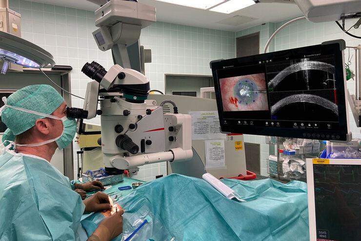
Proveo 8 with intraoperative OCT – a User Evaluation in an University Setting
Optical coherence tomography (OCT) makes structures in the eye visible that lie beneath the surface. When OCT is used intraoperatively, surgeons gain insight into possible pathological changes in the…
How to use a Surgical Microscope as an Operating Room Nurse
Surgical microscopes play an essential role in the modern microsurgery procedures. It provides the surgeon, assistant and operating room staff with a magnified and illuminated high-quality image of…
