Filter articles
Tag
Prodotti
Loading...

Spatial Proteomics Workflow in Blood Cancer (MPNs)
Megakaryocytes play a central role in the biology of myeloproliferative neoplasms (MPNs), yet their in vivo proteomic characterization remains a major challenge due to low abundance and disrupted…
Loading...

AI meets Deep Visual Proteomics (DVP) to Advance Disease Research
In this webinar, Dr. Andreas Mund will introduce a cutting-edge platform that merges Deep Visual Proteomics (DVP) with AI-powered pathology models, enabling high-resolution mapping of key regions in…
Loading...

Exploring Subcellular Spatial Phenotypes with SPARCS
Discover spatially resolved CRISPR screening (SPARCS), a platform for microscopy-based genetic screening for spatial subcellular phenotypes at the human genome scale.
Loading...
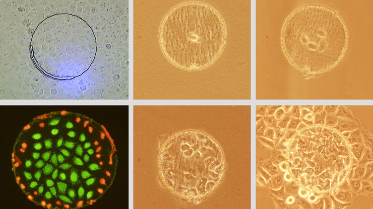
Live Cell Isolation by Laser Microdissection
Laser microdissection is a tool for the isolation of homogenous cell populations from their native niches in tissues to downstream molecular assays. Beside its routine use for fixed tissue sections,…
