Low-input spatial proteomics workflow for megakaryocyte isolation from bone marrow.

Spatial Proteomics Workflow in Blood Cancer (MPNs)

How does spatial proteomics enable low input biomarker discovery in MPN blood cancer?

Low-input spatial proteomics workflow for megakaryocyte isolation from bone marrow. Low-input_spatial_proteomics_workflow_for_megakaryocyte_isolation_from_bone_marrow.jpg

Summary

How can low-input spatial proteomics be used to analyze rare cells in tissue? This webinar presents a validated workflow combining laser microdissection (LMD), AI-based image analysis, and mass spectrometry to profile megakaryocytes directly in bone marrow and uncover disease-specific biomarkers in blood cancer (MPNs).

Using optimized immunohistochemistry and low-input sample preparation, the study achieves robust proteomic profiling from as few as ~100–200 cells. This approach enabled the identification of 4,800+ proteins and revealed, for the first time, distinct disease-specific proteomic signatures across polycythemia vera, essential thrombocythemia, and primary myelofibrosis. The workflow integrates imaging, AI-driven cell selection, and targeted tissue isolation to enable precise and reproducible downstream proteomic analysis.

The findings demonstrate how megakaryocytes actively contribute to disease progression through altered pathways such as extracellular matrix organization, platelet activation, and RNA metabolism. Importantly, the study uncovers potential biomarkers, including senescence-related proteins, which may inform diagnostic strategies and therapeutic development.

For scientists working in hematology, oncology, and spatial biology, this webinar provides a practical and validated workflow to accelerate discovery in complex tissue environments—enabling more precise molecular insights directly from patient samples.

Register now to watch the webinar on demand and gain practical insights you can apply immediately to your research, from sample preparation to spatial data analysis, using real‑world patient samples and proven workflows.

Key learnings

  • How can rare bone marrow cells be profiled directly in situ? Learn a validated spatial proteomics workflow combining AI, LMD, and mass spectrometry.
  • What distinguishes MPN subtypes at the molecular level? Discover disease‑specific megakaryocyte proteomic signatures.
  • How many cells are needed for robust analysis? See how reproducible data is achieved with ~100–200 cells.
  • Which biological pathways are altered in blood cancer? Understand changes in ECM organization, platelet activation, and RNA metabolism.
  • What new opportunities emerge for biomarker discovery? Explore novel candidates, including senescence‑associated proteins.

Megakaryocytes play a central role in the biology of myeloproliferative neoplasms (MPNs), yet their in vivo proteomic characterization remains a major challenge due to low abundance and disrupted tissue architecture. This webinar introduces a novel low-input spatial proteomics workflow that enables, for the first time, direct molecular analysis of these rare cells within their native bone marrow environment.

Why is megakaryocyte proteomics in MPNs challenging?

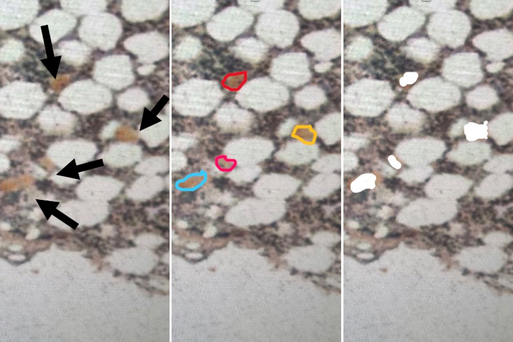
Megakaryocytes are rare, spatially distributed cells embedded in a highly complex and often fibrotic bone marrow environment. Traditional approaches rely on in vitro differentiation or bulk tissue analysis—losing critical spatial and disease specific context.

Key limitations include:

  • Low cell abundance in tissue samples
  • Disrupted bone marrow architecture in MPNs
  • Inadequate sensitivity of conventional proteomic workflows

This creates a major barrier to understanding how these cells drive disease progression.

What new biological insights were uncovered?

This approach revealed, for the first time, that megakaryocytes in MPNs exhibit distinct proteomic signatures across disease subtypes.

Key findings include:

  • Identification of 4,875 proteins across patient cohorts
  • Clear proteomic separation between MPN subtypes
  • Differential pathway regulation:
    • Primary myelofibrosis: ECM organization, RNA metabolism
    • Essential thrombocythemia: platelet activation pathways
    • Polycythemia vera: altered metabolic processes
  • Discovery of novel biomarkers, including senescence-associated proteins

These insights provide a more precise understanding of disease mechanisms at the cellular level.

What does this enable for your research?

This workflow enables researchers to:

  • Generate molecular data from rare cell population
  • Preserve spatial and pathological context in analysis
  • Identify clinically relevant biomarkers and therapeutic targets
  • Reduce experimental iteration through robust, reproducible workflows

Ultimately, it supports faster and more confident translation from biological insight to clinical relevance.

Scroll to top