Confocal Microscopes Articles
 dataset, showing the biochemically distinct structures of a fresh, untreated apple slice.")
How to Prepare Samples for Stimulated Raman Scattering (SRS) imaging
Find here guidelines for how to prepare samples for stimulated Raman scattering (SRS), acquire images, analyze data, and develop suitable workflows. SRS spectroscopic imaging is also known as SRS…

Find Relevant Specimen Details from Overviews
Switch from searching image by image to seeing the full overview of samples quickly and identifying the important specimen details instantly with confocal microscopy. Use that knowledge to set up…

Handbook of Optical Filters for Fluorescence Microscopy
Fluorescence microscopy and other light-based applications require optical filters that have demanding spectral and physical characteristics. Often, these characteristics are application-specific and…
")
Fluorescence Correlation Spectroscopy (FCS)
Fluorescence correlation spectroscopy ( FCS ) measures fluctuations of fluorescence intensity in a sub-femtolitre volume to detect such parameters as the diffusion time, number of molecules or dark…

The Principles of White Light Laser Confocal Microscopy
The perfect light source for confocal microscopes in biomedical applications has sufficient intensity, tunable color and is pulsed for use in lifetime fluorescence. Furthermore, it should offer means…
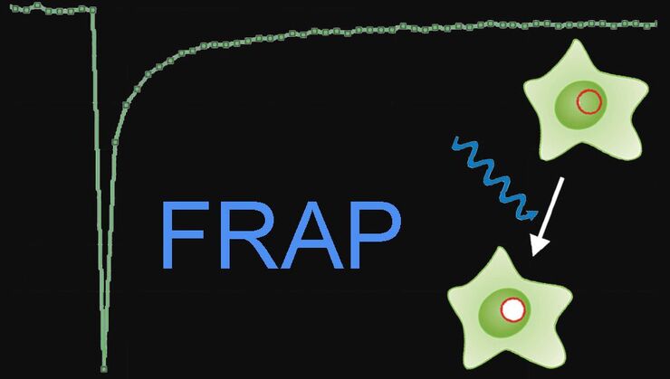
Fluorescence Recovery after Photobleaching (FRAP) and its Offspring
FRAP (Fluorescence recovery after photobleaching) can be used to study cellular protein dynamics: For visualization the protein of interest is fused to a fluorescent protein or a fluorescent dye. A…

Förster Resonance Energy Transfer (FRET)
The Förster Resonance Energy Transfer (FRET) phenomenon offers techniques that allow studies of interactions in dimensions below the optical resolution limit. FRET describes the transfer of the energy…
")
Mosaic Images
Confocal laser scanning microscopes are widely used to create highly resolved 3D images of cells, subcellular structures and even single molecules. Still, an increasing number of scientists are…
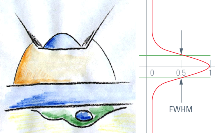
Confocal Optical Section Thickness
Confocal microscopes are employed to optically slice comparably thick samples.
