Contact Us

Christoph Greb , Dr.

Christoph Greb

Christoph Greb studied cell biology, parasitology and virology at the Philipps University in Marburg. In the course of his diploma thesis and his dissertation at the local Institute for Cytobiology and Cytopathology he examined the vesicular transport of apically destined proteins in polarized epithelial cells utilizing biochemistry as well as TIRF and confocal microscopy. From December 2011 he was writing for the Leica Science Lab as a freelancer. After his engagement for Novartis Vaccines & Diagnostics he started as Scientific Writer for the widefield team of Leica Microsystems in October 2013.

Factors to Consider When Selecting a Research Microscope

An optical microscope is often one of the central devices in a life-science research lab. It can be used for various applications which shed light on many scientific questions. Thereby the…

Microscopy in Virology

The coronavirus SARS-CoV-2, causing the Covid-19 disease effects our world in all aspects. Research to find immunization and treatment methods, in other words to fight this virus, gained highest…
Image source: Adobe Stock

How to Sanitize a Microscope

Due to the current coronavirus pandemic, there are a lot of questions regarding decontamination methods of microscopes for safe usage. This informative article summarizes general decontamination…

Improving RNA Analysis with Laser Microdissection

Parkinson’s disease is a progressive neurodegenerative disorder connected with cell death of dopamine-releasing neurons in the brain. Differences in gene expression between individual…

How to improve your Alzheimer Protein Analysis with Laser Microdissection

Brain Research: Collect pure starting material for proteomics - Improve your workflow with Laser Microdissection - Many brain diseases result from protein malfunction, misfolding and agglutination.…

How to improve your DNA Mutation Analysis Workflow with Laser Microdissection

DNA mutations lead to abnormal proteins or missing functional proteins, which can cause cells to multiply uncontrollably and become cancerous. To find and understand the underlying mutation for a…
Mammalian cell culure. Phase contrast and fluorescence image.

Introduction to Mammalian Cell Culture

Mammalian cell culture is one of the basic pillars of life sciences. Without the ability to grow cells in the lab, the fast progress in disciplines like cell biology, immunology, or cancer research…
Convalaria

Introduction to Widefield Microscopy

This article gives an introduction to widefield microscopy, one of the most basic and commonly used microscopy techniques. It also shows the basic differences between widefield and confocal…
Dronpa chromophore

Photoactivatable, Photoconvertible, and Photoswitchable Fluorescent Proteins

Fluorescent proteins (FPs) such as GFP, YFP or DsRed are powerful tools to visualize cellular components in living cells. Nevertheless, there are circumstances when classical FPs reach their limits.…
Left: Tissue cells marked with an immunolabel (FITC) illuminated with wide-band UV excitation. Note the tissue structure with blue autofluorescence. Right: Same tissue and same immunostaining with FITC label illuminated with epi-illumination using narrow-band blue (490 nm) light. Note the increased image contrast (Ploem, 1967)

Milestones in Incident Light Fluorescence Microscopy

Since the middle of the last century, fluorescence microscopy developed into a bio scientific tool with one of the biggest impacts on our understanding of life. Watching cells and proteins with the…
HeLa cells stimulated with LPS. Image has been subjected to deconvolution.

Chronic Inflammation Under the Microscope

In the course of chronic inflammation certain body areas are recurrently inflamed. This goes along with many human diseases. With the help of widefield light microscopy, the underlying processes can…
Gene editing with Cas9

Gene Editing with CRISPR/Cas9 - Breakthrough in Genome Engineering

The CRISPR/Cas9 system is one of several different bacterial systems for defense against viral attacks. It consists of two main components. One is a small piece of RNA which binds to the viral target…
Infinity port

Infinity Optical Systems

“Infinity Optics” refers to the concept of a beam path with parallel rays between the objective and the tube lens of a microscope. Flat optical components can be brought into this “Infinity Space”…

Universal PAINT – Dynamic Super-Resolution Microscopy

Super-resolution microscopy techniques have revolutionized biology for the last ten years. With their help cellular components can now be visualized at the size of a protein. Nevertheless, imaging…

Sample Preparation for GSDIM Localization Microscopy – Protocols and Tips

The widefield super-resolution technique GSDIM (Ground State Depletion followed by individual molecule return) is a localization microscopy technique that is capable of resolving details as small as…

Image Processing for Widefield Microscopy

Fluorescence microscopy is a modern and steadily evolving tool to bring light to current cell biological questions. With the help of fluorescent proteins or dyes it is possible to make discrete…

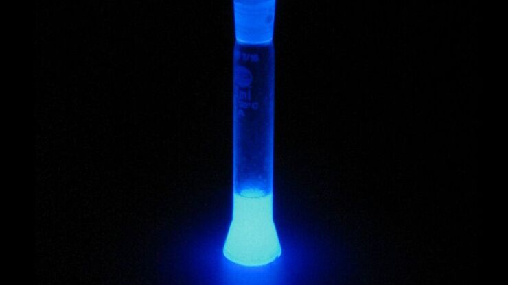
Basic Principles of Luminescence

There are a lot of light-emitting processes occurring in nature. Luminescence is an umbrella term for those kinds of events where light emission is not the result of high temperatures. This article…
Jellyfish Aequorea Victoria

Fluorescent Proteins - From the Beginnings to the Nobel Prize

Fluorescent proteins are the fundament of recent fluorescence microscopy and its modern applications. Their discovery and consequent development was one of the most exciting innovations for life…
Scroll to top