Contact Us

Thomas Veitinger , Dr.

Thomas Veitinger

Dr. Thomas Veitinger studied Biology at the Ruhr-University Bochum, Germany and received a diploma in Developmental Neurobiology. In his Ph.D. thesis at the Department of Cell Physiology of the Ruhr-University in Bochum, he investigated odor-driven subcellular calcium dynamics of human spermatozoa and gained experience in live-cell imaging methods and general physiology. As a post-doc, he worked in the department for chemosensation of the RWTH Aachen University, Germany lead by Prof. Dr. Marc Spehr. There, he investigated male germ stem cells using the patch-clamp technique and live-cell imaging. Since May 2011, he is employed by Leica Microsystems as an Application Manager for live-cell imaging, electrophysiology and neuroscience applications.
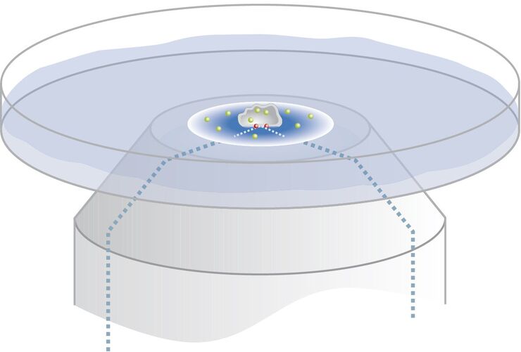
Controlling the TIRF Penetration Depth is Mandatory for Reproducible Results

The main feature of total internal reflection fluorescence (TIRF) microscopy is the employment of an evanescent wave for the excitation of fluorophores instead of using direct light. A property of the…
Primary leaves of cowpea (Vigna unguiculata "California Blackeye") inoculated with cowpea mosaic virus (CPMV) containing the GFP-gene inserted between the movement protein (MP) and the capsid proteins (CPs) in the viral RNA 2

Introduction to Live-Cell Imaging

The understanding of complex and fast cellular dynamics is an important step to get insight into biological processes. Therefore, today’s life science research more and more demands studying…
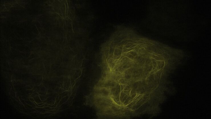
TIRF Image of Tubulin, YFP, penetration depth: 120 mm

Applications of TIRF Microscopy in Life Science Research

The special feature of TIRF microscopy is the employment of an evanescent field for fluorophore excitation. Unlike standard widefield fluorescence illumination procedures with arc lamps, LEDs or…
Snapshot from a time lapse of a calcium imaging experiment using the ratiometric calcium indicator Fura-2.

Ratiometric Imaging

Many fundamental functions of a cell strongly depend on delicate, but nevertheless dynamic balances of ions (e.g. calcium, magnesium), voltage potentials and pH between the cell’s cytosol and the…
Modulation contrast visualizes transparent, low-contrast specimens.

Integrated Modulation Contrast (IMC)

Hoffman modulation contrast has established itself as a standard for the observation of unstained, low-contrast biological specimens. The integration of the modulator in the beam path of themodern…
Scroll to top