
Cryo Electron Tomography Workflow
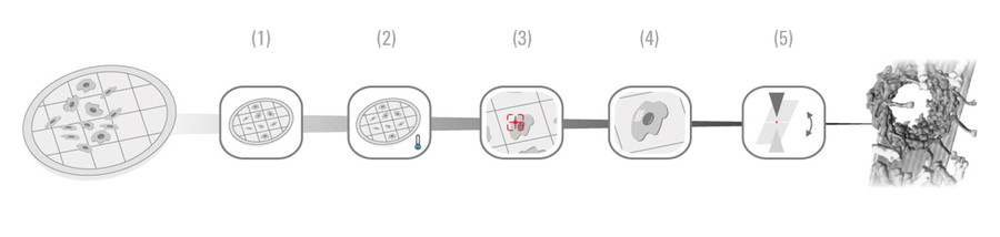
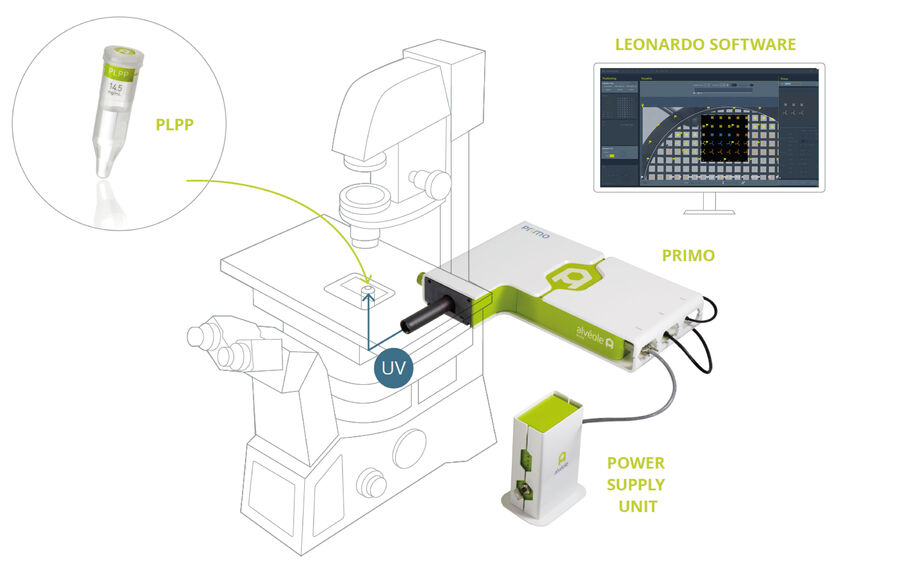
Step 1: Micropatterning with PRIMO
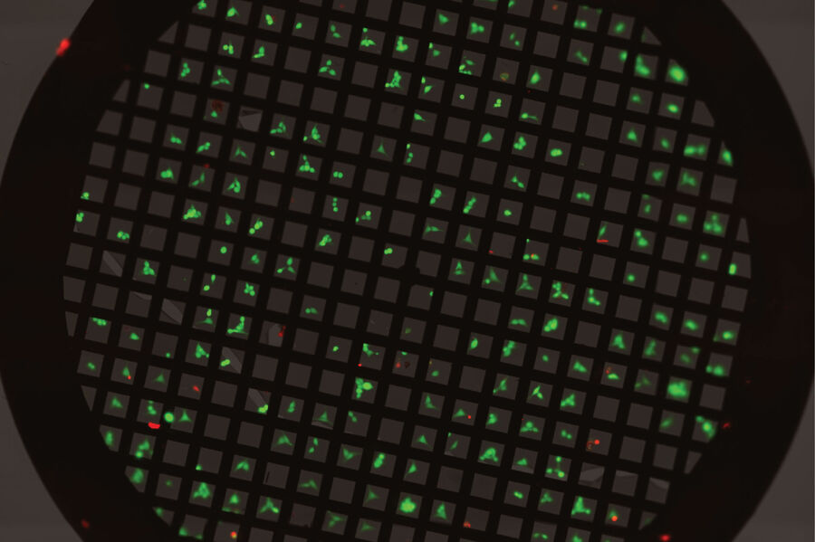
Overcome the first challenges of the cryo-ET sample preparation process by using PRIMO, a micropatterning system that allows control of cell adhesion, spreading and shape on EM grids. It enables precise cell positioning and optimized cell spreading, while leaving the surface of the EM grid undamaged. The software automatically aligns your patterns within the mesh of your EM grid, ensuring that your cells are optimally positioned for further processing.
Learn more about PRIMO: https://www.alveolelab.com/our-products/primo-micropatterning/
Step 2: Vitrification
Prior to freezing, the sample is maintained in an humidity-controlled environmental chamber at a stable temperature.
The sample is then vitrified by plunge freezing with the automatic plunge freezer EM GP2. The vitrification process inhibits ice crystal formation ensuring the cellular content is as close to the native state as possible.

Step 3: Selection
For an efficient workflow, suitable cells and target regions have to be preselected. This is achieved by using the cryo light microscope THUNDER Imager EM Cryo CLEM. The sample can then be transferred to the Thermo Scientific™ Aquilos™ for milling. Full connectivity between the instruments means that the defined region and coordinates are securely and precisely transferred for immediate relocation by the Aquilos. No need to invest time searching for the relevant target positions!
To avoid contamination of the precious specimen, the Integrated Cryo Tomography workflow ensures a secure transfer of the sample between the systems involved. The dedicated cartridge system safeguards your sample throughout the workflow providing the solid fundament for your reliable scientific results.
(Thermo Scientific, Krios and Aquilos are Trademarks by Thermo Fisher Scientific.)

Step 4: Milling
After preselection and targeting in the THUNDER Imager EM Cryo CLEM, the sample is transferred to the Thermo Scientific Aquilos, a dedicated Cryo DualBeam electron microscope.
In the past, it was impossible to resolve the interior of cells at subnanometer resolution as many samples were simply too thick to be imaged by cryo-electron tomography.
The Aquilos overcomes the thickness limitations by employing a scanning electron beam (SEM) and a focused ion beam (FIB). While the electron beam is used for imaging, a beam of gallium ions ensures the precise milling of vitrified cells.
By the milling process, a thin ice sheet, the on-grid lamella is created and which can then be examined using electron tomography.
 and the focused ion beam (FIB) for milling a thin ice sheet into the cell.")
Step 5: 3D Cryo Tomography
The EM grid that carries the cells including the on-grid lamella, is transferred from the Aquilos to the Thermo Scientific Krios™ G3i (Cryo TEM).
The Cryo TEM images the area of interest multiple times from different angles by tilting the sample incrementally.
The single images are computationally aligned and reconstructed to generate a three-dimensional tomogram from the lamella and its content in subnanometer resolution.

Related Articles
-
 labeled with membrane-permeable calcein, high-pressure frozen in salt water using EM ICE.")
High-Pressure Freezing for Organoids: Cryo CLEM & FIB Lift Out
Master cryo EM workflow steps for challenging 3D samples: when to choose HPF vs. plunge freezing,…
Jan 22, 2026Read article -

Integrated Serial Sectioning and Cryo-EM Workflows for 3D Biological Imaging
This on-demand webinar explores how integrated tools can support electron microscopy workflows from…
Jul 22, 2025Read article -

Revealing Sodium Battery Degradation via Cryo-EM and CryoFIB
Explore how cryogenic electron microscopy and focused ion beam techniques uncover the intrinsic…
Jul 22, 2025Read article
