
Key Learnings
What to expect from the poster
- Learn how Cell DIVE multiplexed imaging can be used to explore human brain samples
- See how antibodies from Cell Signaling Technology can accelerate multiplexed imaging workflow
- Understand how cell identity and spatial heterogeneity of the brain tissue contribute to the structure of the Alzheimer’s brain
About Alzheimer's disease
Alzheimer's disease (AD) is the most common type of dementia causing cognitive impairment in midlife and late life. It is a neurodegenerative disorder with both genetic and non-genetic causes. AD is characterized by the presence of β-amyloid-containing plaques and phosphorylated tau neurofibrillary tangles. Current treatments for AD are limited, and researchers are investigating the role of histopathological features for potential therapeutic approaches. The Cell DIVE Multiplexed Imaging Solution, along with CST's antibodies, provides a way to study synaptic processes and identify cell types around AD hallmarks.
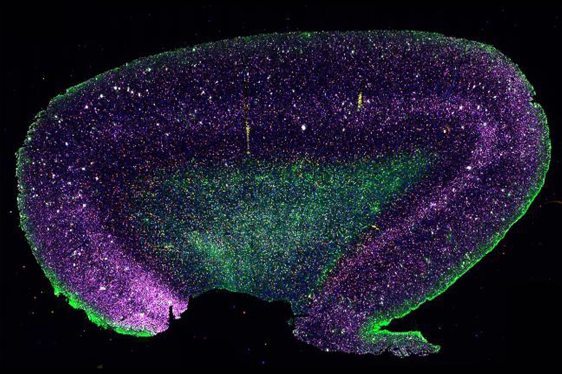
The study
This approach helps to recognize disease-related microglia and comprehend cellular relationships that contribute to AD. This method identifies spatially co-localized cell populations and serves as a useful tool for exploring AD. CST's many antibodies can help spot changes in the way cells communicate and different cell types in diseased tissue. With a novel CST panel, researchers can examine the protein landscape and look at how Amyloid-β and tau are expressed in people with Alzheimer's.
CST's antibodies are subjected to rigorous validation, so they work well on tissue that has been preserved with formalin and buried in paraffin. The study's direct connections were repeatedly colored, giving a description of the spatial makeup of AD brain tissue. The tissue-preserving nature of Cell DIVE enables deeper investigation, presenting a new technique for neuroscience to understand the differences in space in the human brain and their connection to illness. Targeted studies of certain brain regions, such as the amygdala, could reveal fresh spatial features linked to AD, creating new paths for future therapeutic approaches.


.")
