Key Learnings
Dr. Bach and her team use the Leica PROvido surgical microscope, a multidisciplinary microscope allowing to see more of the surgical site in one view. It provides a bright and fully-focused view deeper into narrow cavities. The PROvido operating microscope unites premium surgical optics with a responsive and stable floor stand.
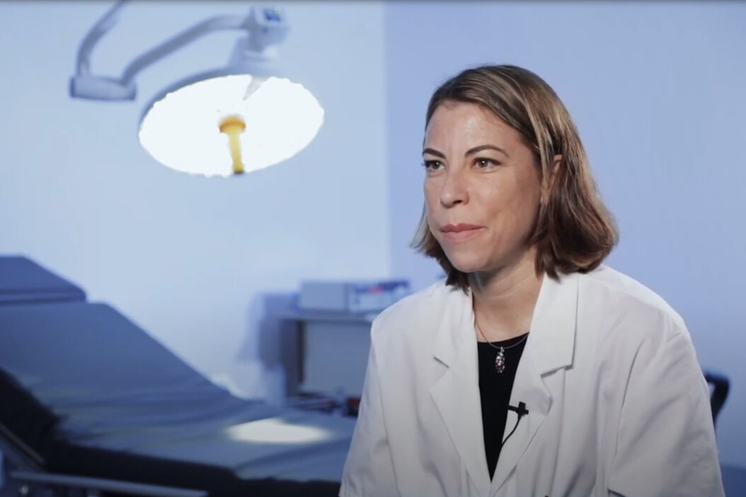
In the below video, Dr. Bach explains the benefits of PROvido in plastic reconstructive surgery including ease of use, a good distribution of light and low heat generation. The microscope also allows the surgeon and his assistant to operate in a comfortable position. And all the OR staff can see the images, supporting communications and teaching. For the scrub nurse, the microscope is easy to drape.
Discover Dr. Bach’s video interview below as well as a full transcript in English. To learn more about PROvido or find the right plastic surgery microscope for your needs, contact a Leica representative.
About the PROvido microscope
The PROvido surgical microscope provides unique benefits for plastic reconstructive surgery. It features outstanding optics and an extended depth of field to see everything you need to see. There is no need to search for vital details or constantly refocus. The innovative light management system protects patient skin and tissue. In addition, the PROvido microscope stand is extremely lightweight and versatile for efficient set-up and workflow in the OR. The FL800 ULT intraoperative video angiography module allows to observe blood flow in real time. Used in conjunction with ICG fluorescent agent, it enables visual assessment of vessel patency during vascular procedures.
To learn more about PROvido or find the right plastic surgery microscope for your needs, contact a Leica representative.
The statements of the healthcare professional included in this article reflect only her opinion and personal experience. They do not necessarily reflect the opinion of any institution with whom she is affiliated.
Transcription
“From the first time I used this Leica microscope, it gave me a good impression. It is easy to set-up and the microscope is very light to use.
I’m Dr. Christine Bach and I’m a plastic surgeon specializing in head and neck surgery. I carry out reconstructive surgery for patients with ENT cancers by microsurgery.
Reconstructive surgery must be both functional and aesthetic. Functional because we are working with the upper aerodigestive tract and the patient must be able to talk, eat and breathe and we have to ensure a 3D reconstruction because we are working with bone, muscle, mucosa and the teeth. And just as importantly you have the aesthetics, this is because you are working on the face.
All of this makes reconstructive surgery very rewarding as I am trying to make a reconstruction that is as close to the original as possible. One thing that isn’t taken into account is that after performing microsurgery, your eyes are often sore and the team suffers from headaches. Here, this wasn’t the case. We used, I think, just 18% of the light, yet the operating field of view was good.
The thing I liked the best was that the microscope didn’t generate heat and therefore the flap didn’t heat up during the operation. With other microscopes, you have to apply compresses soaked in physiological serum and I believe the flap can suffer from that. Here it’s really an interesting advantage.
The head of the microscope is very easy to move and position. With the optics, especially for the surgeon assistant, it is simple to set and to incline, even for neck surgery, and finally this results in very comfortable positioning for all.
The buttons are easy to use: the zoom, the position changer. The camera used for viewing indocyanine green fluorescence was also easy to set up. During a particularly delicate operation today with difficult microvascular anastomoses, we felt well supported for this type of operation.
The operator and the other theater staff saw exactly what was happening with an image quality that was very close to what could be seen in the microscope, which is very useful. The possibility to take film and to take photos is also very interesting for teaching purposes.
Even the scrub nurse says that the cover was great. No but it’s important! [Comment from nurse: Yes, but it’s true, since you have never covered it].
The set-up and the fact that the microscope is easy to position, to adjust, is important and it’s true, when it is badly balanced, you press the button and the head of the microscope starts rocking, it is always, difficult. Here it’s easy to adjust, it’s something that does everything on its own, it’s true that it’s great.”
Related Articles
-

The Guide to Augmented Reality in Microsurgery
In an era of technological advancement, Augmented Reality (AR) is rapidly transforming the medical…
Jun 16, 2025Read article -

Using GLOW800 AR in Radial Forearm Flap Free Phalloplasty
In this video, Chief Microsurgeon Professor Küntscher and his team perform a radial forearm free…
Apr 04, 2022Read article -

How AR Surgery Benefits Radial Forearm Free Flap Phalloplasty
The goal of penile reconstruction is to provide an aesthetic penoid with tactile and erogenous…
Mar 14, 2022Read article
