Contact Us

Utility of Intraoperative OCT in Sub-Retinal Gene Therapy

Discover a case study on the use of intraoperative Optical Coherence Tomography (OCT) for pediatric gene therapy

The intraoperative OCT showed the ellipsoid had separated from the inner retina with focal attachment to the retinal pigment epithelium (RPE). Images provided by Mr. Robert Henderson Ellipsoid_separated_from_the_inner_retina_with_focal_attachment_to_the_retinal_pigment_epithelium.jpg

The RPE65 gene provides instructions for making a protein that is essential for normal vision [1]. Mutations in the RPE65 gene lead to reduced or absent levels of RPE65 activity, blocking the visual cycle and resulting in impaired vision [2].

Gene therapy treatment, which for this gene is delivered via a surgical injection under the retina, gives patients a normal copy of the RPE65 gene. In delivering a correct copy of the gene, sight is improved and the progress of the dystrophy halted [3].

About the case study

Key Learnings:

  • Learn about the surgical management of a 3-year-old girl with the classic features of RPE65 disease: an early onset severe retinal dystrophy including light staring and nyctalopia
  • Discover the different steps of subretinal gene therapy injection
  • Understand the role of intraoperative OCT, particular for bleb placement

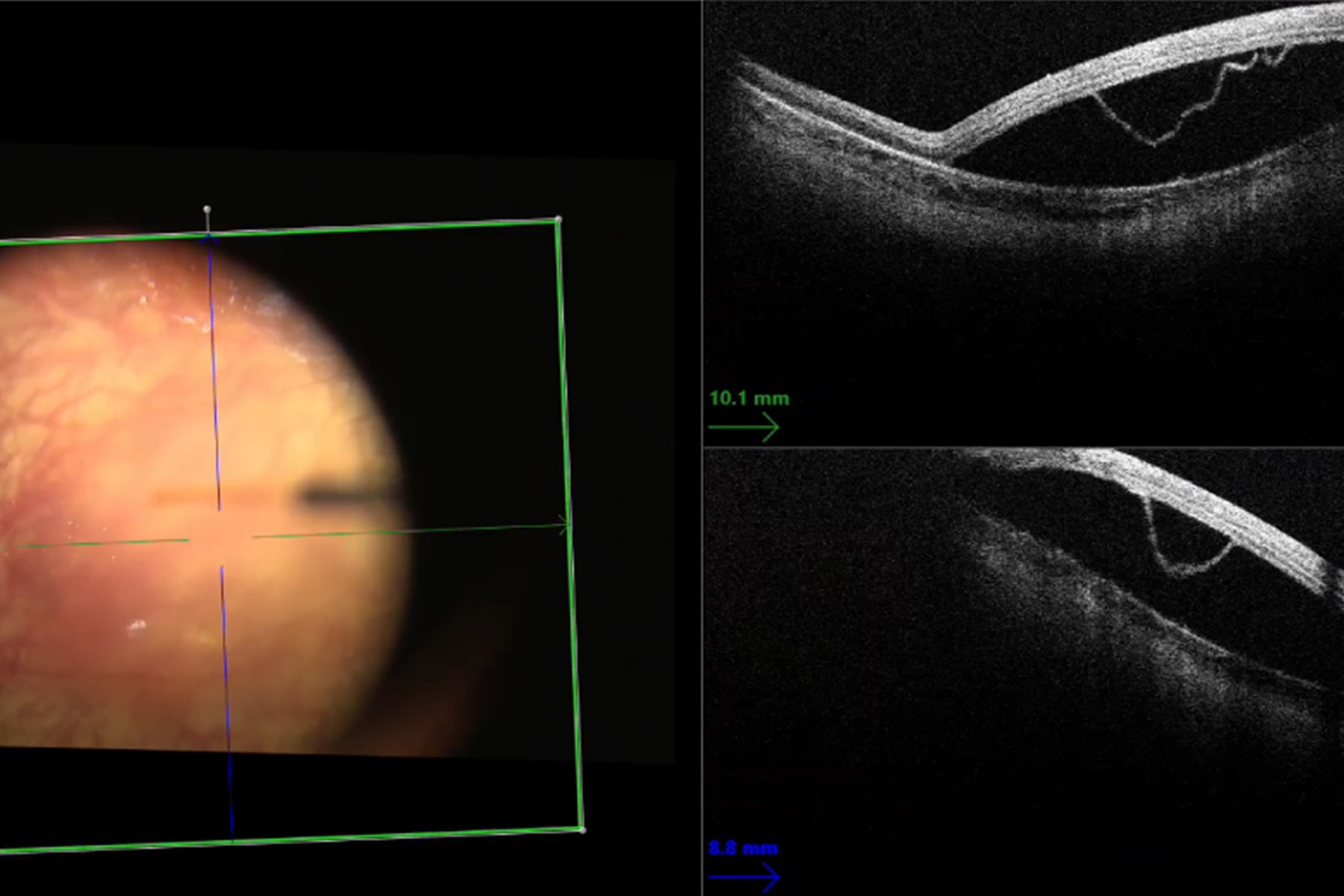
Gene therapy surgical procedures require a high level of precision and pose specific challenges in pediatric patients. The use of intraoperative Optical Coherence Tomography (OCT) is essential to ensure correct bleb placement and manage potential complications.

Case Description

A 3-year-old Caucasian girl presented with the classic features of RPE65 disease: an early onset severe retinal dystrophy including light staring and nyctalopia. She was diagnosed with compound heterozygous mutations in the RPE65 gene. She had nystagmus, poor low illumination visual function and reduced photopic acuity.

Pre-operative assessment

The patient’s vision was 1.2 LogMAR and she was myopic. The pre-operative retinal fundus imaging showed mild vascular attenuation, normal macula volume, an intact ellipsoid, and no obvious dystrophy features. She had an absent electroretinogram (ERG) and her pattern visual evoked potential (pVEP) was significantly reduced.

Continue Reading 

Want to discover how Mr. Henderson injected gene therapy under the retina utilizing intraoperative OCT on the Proveo 8 surgical microscope?

Complete the form to download the full case study. 

Download now!

By clicking on the SUBMIT button, I confirm that I have reviewed and agree with Leica Microsystems GmbH Terms of Use and their Privacy Policy. I also understand my privacy choices as they pertain to my personal data, as detailed in the aforementioned Privacy Policy under ‘’Your Privacy Choices’’.

Please note that off-label uses of products may be discussed. Please check with regulatory affairs for cleared indications for use in your region. The statements of the healthcare professionals included in this clinical case reflect only their opinion and personal experience and not those of Leica Microsystems. They also do not necessarily reflect the opinion of any institution with whom they are affiliated.

Background image
Scroll to top