
Birefringence is the key to polarization microscopy
Birefringent materials are optically anisotropic and the refractive index of such materials depends on the direction in which light propagates through it. Birefringence means a ray of unpolarized light is divided into two rays by refraction when passing through the material. Birefringent materials have a highly ordered molecular structure, like crystals of calcite or boron nitride. Biological specimens also can be birefringent, for example cellulose and starch. In combination with linearly polarized light, birefringence can be used in microscopy to achieve interference of the two resulting rays, which can produce color effects like rings and the highlighting of structures.
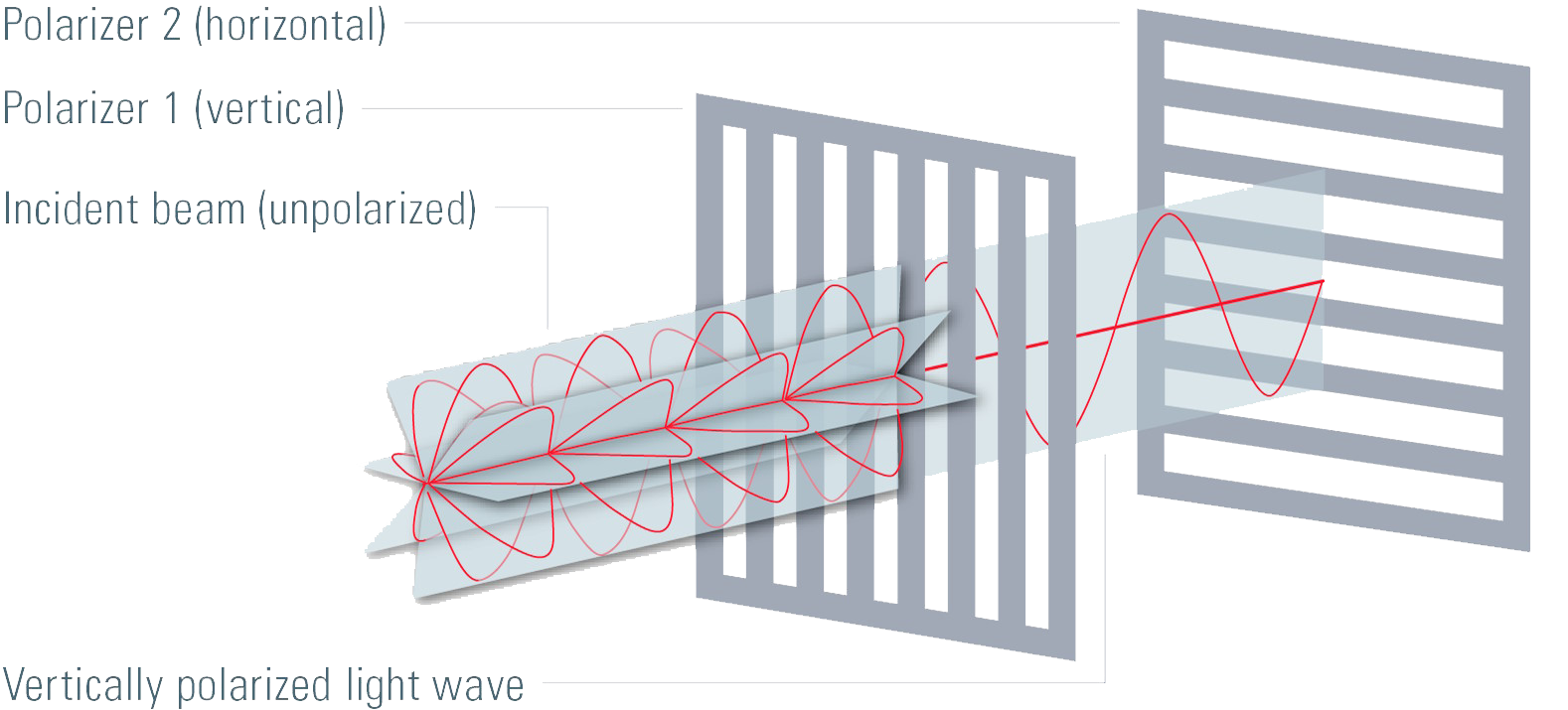
Alignment and beam path of a polarizing microscope
A conventional optical microscope needs at least two additional components to perform polarized light microscopy. For detection of birefringence, linearly polarized light must be used for illumination of the specimen or sample. Therefore, two polarizing filters have to be inserted into the beam path of the microscope. The first polarizing filter produces polarized light which illuminates the specimen and the second polarizing filter, called the analyzer, restricts the passage of light which is detected to refracted light with a specific polarization.
The transmission axes of the polarizing filters have to be crossed at an angle of 90°, meaning the extinction axis of one filter is aligned with the transmission of the other, to achieve the so called “dark position”. When the polarizing filters are set at this position, no light will pass to the camera or eyepieces and the image will be dark. Setting up the dark position is an important step for polarized light microscopy as it ensures that only light which experiences a change in polarization after passing through the specimen will be visible.

Polarizer and analyzer
When light passes through the first polarizing filter, it is linearly polarized. If some of the linearly polarized light passes through the correct polarization plane of a birefringent material, it is refracted, split into two rays, and the polarization plane of these rays is turned by 90°. The refracted rays then pass through the second polarizer (analyzer), if it is aligned correctly (i.e., 90° relative to the first polarizing filter). Therefore, for this setup only birefringent specimens or materials produce an image when observed with a polarized light microscope.
It is important that the polarization axis of the birefringent material being observed is the same as that of the polarized light produced by the first polarizer filter. For this reason, many polarization microscopes are equipped with a rotating stage to ensure easy alignment of the sample’s polarization plane with the one of the first polarizer. Various accessories are available for special applications where polarization microscopy is useful.
A Bertrand lens is used in the microscope for conoscopic observation of interference patterns from crystals where the light rays are focused in the plane of the objective rear aperture [1,2]. Additionally, a retardation plate or compensator is useful for quantitative analysis of birefringent specimens.
Applications for polarization microscopes


 alloy etched with Barker’s reagent taken with polarization microscopy.")