Contact Us

Cancer Research

Cancer is a complex and heterogeneous disease caused by cells deficient in growth regulation. Genetic and epigenetic changes in one or a group of cells disrupt normal function and result in autonomous, uncontrolled cell growth and proliferation.

Imaging has become a key tool in the study of cancer biology. High-resolution imaging is indispensable for the study of genetic and cell signaling changes that underlie cancer, whereas live-cell imaging is crucial for a deeper understanding of function and disease mechanisms. Microscopy techniques are also essential for the study of spatial relationships between different types of tumor cells. They are also important for understanding the role of the immune system in battling cancerous cells. For the latter, researchers rely on multicolor imaging for a faster rate of discovery.

Simply get in touch!

Our experts on solutions for Cancer Research are happy to help you with their advice.

Challenges when using imaging to study cancer

Optimal temporal and spatial resolution

Research into cancer therapeutics often requires the combination of fluorescence microscopy and innovative functional assays. With optimal temporal and spatial resolution, researchers are able to monitor dynamic events in living cells, such as cell migration and metastasis. These dynamic processes are at the core of cancer development.

Visualizing in real time

Understanding these processes has been challenging due to the difficulty of visualizing tumor cell behavior in real time. Fast imaging over prolonged periods of time tends to come with a sacrifice: either decreased resolution or, more often, harm to your precious specimens. The challenge is finding the imaging technique and system that provides you with the best data with the highest resolution while keeping the cells alive so that you can follow the processes of interest.

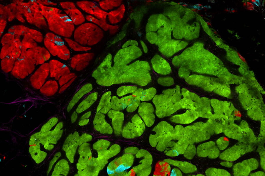
Multiplexing to understand mechanisms of disease

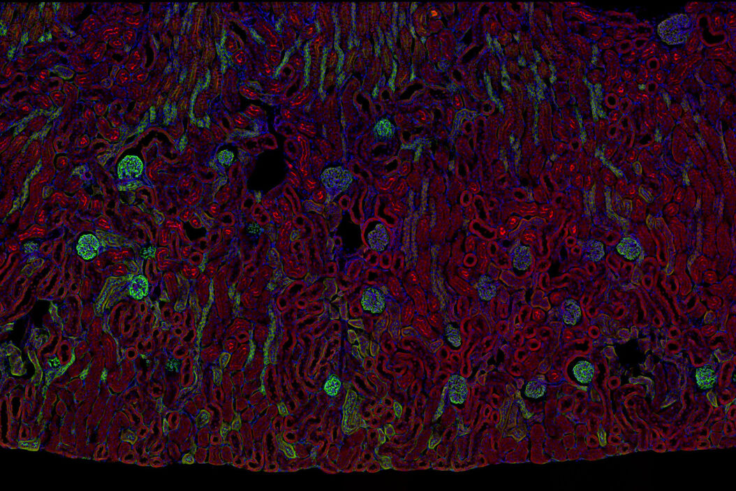
Multicolor fluorescence microscopy, either confocal or widefield based, is a fundamental tool to understand the spatial context, co-localization, and proximity of multiple biomarkers when studying complex events, such as immunosuppression or angiogenesis. This aim can often be challenging, as there are limits to the number of fluorophores you can successfully distinguish with this “multiplexing” approach.

Fortunately, there are innovative imaging systems and strategies to improve the separation of fluorophores (e.g. FluoSync - a streamlined approach for simultaneous multiplex fluorescent imaging using a single exposure) and increase the number of fluorescent probes to that which is needed in your experiment.

Finding the right tools

Cancer is complex and requires a myriad of methods that include spatiotemporally resolved, live-specimen, and single-cell imaging. More insights into cellular processes concerning cancer will come likely from methods with the highest possible resolution and multiparametric image analysis. Approaches like fluorescence confocal microscopy enable you to study multiple targets within tissues or cellular structures.

Advanced imaging techniques, such as super-resolution or, more recently, lifetime imaging or lightsheet, help you to better understand the molecular interactions and regulatory mechanisms behind tumor initiation, progression, and response to treatment.

Laser microdissection or correlative light and electron microscopy (CLEM) enable the study of spatial receptor arrangements in membranes and genome organization in cell nuclei.

related tools

Super-Resolution Microscopy

Super-resolution light microscopy empowers you to study subcellular structures and dynamics with greater detail. While the spatial resolution of confocal image acquisition is doubled with the LIGHTNING technology, when using STED it can deliver insights at the nanoscale. Learn more here about Leica super-resolution methods and how they enable novel discoveries in the fields of virology, immunology, neuroscience, and cancer research.

Fluorescence Microscopy Solutions

Find out how fluorescence microscopes from Leica Microsystems support your research. Fluorescence is one of the most commonly used physical phenomena in biological and analytical microscopy for its high sensitivity and high specificity. Fluorescence is a form of luminescence that through microscopy allows users to determine the distribution of a single molecule species, its amount and its localization inside a cell.

Fluorescence Lifetime Imaging Solutions

Leica Microsystems is at the cutting edge of today’s fluorescence lifetime imaging advances. Our systems make lifetime imaging faster and easier to use than ever before, bringing its advantages to every day confocal imaging experiments.

Multiplex Imaging Reveals Survival Markers after Cancer Care

Colorectal cancer is a high incidence and high mortality cancer. Currently, postoperative chemotherapy benefits only a minority of patients, and thus, new tools are necessary to screen patients and identify those at increased risk. Tissue samples from hundreds of patients were analyzed using Cell DIVE’s multiplex imaging to reveal the fine cellular determinants of survival following cancer treatment. 

Read More

Tumor MHC Expression and Intralesional IL2 Response in Melanoma

Genomics profiling and Cell DIVE multiplex imaging allows researchers to understand the immune cell phenotypes that most strongly predict response to IL2 immunotherapy in melanoma patients suffering metastasis.

Read More

Hyperplex Cancer Tissue Analysis at Single Cell Level with Cell DIVE

The ability to study how lymphoma cell heterogeneity is influenced by the cells’ response to their microenvironment, especially at the mutational, transcriptomic, and protein levels. Protein expression studies offer the most relevant information about the nature of cellular interactions and protein expression levels. A hyperplexed workflow can be applied for studying multiple proteins from the same cancer tissue.

Read More

Related Articles

Read our latest articles about Cancer Research

The knowledge portal of Leica Microsystems offers scientific research and teaching material on the subjects of microscopy. The content is designed to support beginners, experienced practitioners and scientists alike in their everyday work and experiments.

More Articles
Spheroid stained with Cyan: Dapi nuclear countertain; Green AF488 Involucrin; Orange AF55 Phalloidin Actin; Magenta AF647 CK14.

Notable AI-based Solutions for Phenotypic Drug Screening

Learn about notable optical microscope solutions for phenotypic drug screening using 3D-cell culture, both planning and execution, from this free, on-demand webinar.
Hepatocellular Carcinoma with 13 biomarkers shown – Beta-Catenin, CD3D, CD4, CD8a, CD31, CD44, CD163, DAPI, PanCK, PCK26, PD1, SMA, and Vimentin.

Understanding Tumor Heterogeneity with Protein Marker Imaging

Explore tumor heterogeneity and immune cell dynamics. See how quantitative imaging analysis reveals spatial relationships and molecular insights crucial for advancing cancer research and therapeutics.
Esophageal tissue with a squamous cell carcinoma labelled with the 4 biomarkers PanCk, DAPI, NaKATPase, and Vimentin.

Discover how Multiplexed Bioimaging can Advance Cancer Research

Explore multiplexing with up to 60 biomarkers, enabling advanced tumor imaging approaches to gather precise, spatially-resolved single-cell data that helps enhance cancer research and clinical…
Multi-tissue array with 4 markers shown including DAPI, NaKATPase, PanCk, and Vimentin.

Spatial Biology: Learning the Landscape

Spatial Biology: Understanding the organization and interaction of molecules, cells, and tissues in their native spatial context
Branched organoid growing in collagen where the Nuclei are labeled blue. To detect the mechanosignaling process, the YAP1 is labeled green.

Examining Developmental Processes In Cancer Organoids

Interview: Prof. Bausch and Dr. Pastucha, Technical University of Munich, discuss using microscopy to study development of organoids, stem cells, and other relevant disease models for biomedical…
Cell counts for each biomarker were divided by total number of cells to give a percentage of biomarker positive cells out of total cells for each biomarker.

Methods to Improve Reproducibility in Spatial Biology Research

Establish reproducibility results for a Cell DIVE multiplexed imaging study in cancer research using the BAB 200 automated system from ASLS and validated antibodies from CST
PDAC Multiplexed imaging of CST panels enables an examination of immune cell components in pancreatic ductal adenocarcinoma (IPDAC) tissue on a single slide.

Characterizing tumor environment to reveal insights and spatial resolution

Antibodies from Cell Signaling Technology are validated for use with the Cell DIVE multiplexing workflow and used to probe cell lineages in the tumor microenvironment
Cancer cells

The Role of Iron Metabolism in Cancer Progression

Iron metabolism plays a role in cancer development and progression, and modulates the immune response. Understanding how iron influences cancer and the immune system can aid the development of new…
Raw widefield (left) and Computationally Cleared image (right) of mouse neuromuscular junctions acquired with a THUNDER Imager. Courtesy of A. Yung and M. Krasnow in California, USA.

Neural Basis of Breathing, Vocalization, and Diseases of the Larynx

This article shows how research on diseases of the larynx can be improved with a THUNDER Imager and Computational Clearing. Motor neurons in whole mounts of mouse laryngeal muscle are better…
Background image
Scroll to top