Super-resolution image of intracellular structures using a STELLARIS STED microscope and TauSTED Xtend. The magenta color shows actin filaments stained with ATTO 647N.

Cellular Analysis

Cellular analysis helps researchers gain deeper insight into cellular functions and subcellular structures. Advanced microscopy imaging solutions and AI-powered analysis software from Leica Microsystems enable precise functional and correlative imaging, as well as analysis of single cells, live cells, and endpoint assays.

Simply get in touch!

Our experts on solutions for cellular analysis applications are happy to help you with their advice.

Mica Microhub No constraints - 4x more data with 100% correlation

How do cellular mechanisms influence disease progression?

Understanding cellular mechanisms is vital for fields like cancer research and Alzheimer's disease. For example, cellular signaling pathways regulate cancer cell proliferation, while distinct cellular alterations contribute to Alzheimer's pathology.

What are the optimal methods for live cell analysis?

Advanced imaging methods – including widefield, confocal, multiphoton, and super-resolution microscopy – enable real-time observation of live cells. These techniques offer precise insights into cellular dynamics and behavior, supporting diverse research in cellular analysis.

How can comprehensive cellular analysis be achieved?

Integrating diverse imaging techniques, such as functional and correlative imaging, enables a more thorough analysis. This approach offers deeper insights into cellular processes and interactions.

How can Leica solutions help researchers studying cellular analysis?


High-resolution imaging

For precise analysis and accurate interpretation, Leica instruments, including optical compound and confocal microscopes, offer high-resolution imaging. This enables researchers to visualize fine subcellular details with clarity. 


AI-powered analysis software

Researchers using the AI-powered analysis software from Leica Microsystems, such as Aivia, enhance their ability to analyze complex data. Dedicated analysis tools for various fields, like cell biology and neuroscience, are available with automated features that save time and improve the accuracy of results, even for complex experiments.


Versatile imaging solutions

Ranging from functional to correlative imaging, the versatility of Leica microscopy solutions ensures that researchers have the right tools tailored to their specific scientific applications and experimental goals.

Related Articles

Read our latest articles about Cellular Analysis

The knowledge portal of Leica Microsystems offers scientific research and teaching material on the subjects of microscopy. The content is designed to support beginners, experienced practitioners and scientists alike in their everyday work and experiments.

More Articles
AI-based cell counting performed with a phase-contrast and fluorescence image using the Mateo FL microscope.

Precision and Efficiency with AI-Enhanced Cell Counting

This article describes the use of artificial intelligence (AI) for precise and efficient cell counting. Accurate cell counting is important for research with 2D cell cultures, e.g., cellular dynamics,…

Live-Cell Imaging Techniques

The understanding of complex and/or fast cellular dynamics is an important step for exploring biological processes. Therefore, today’s life science research is increasingly focused on dynamic…
Virally labeled neurons (red) 
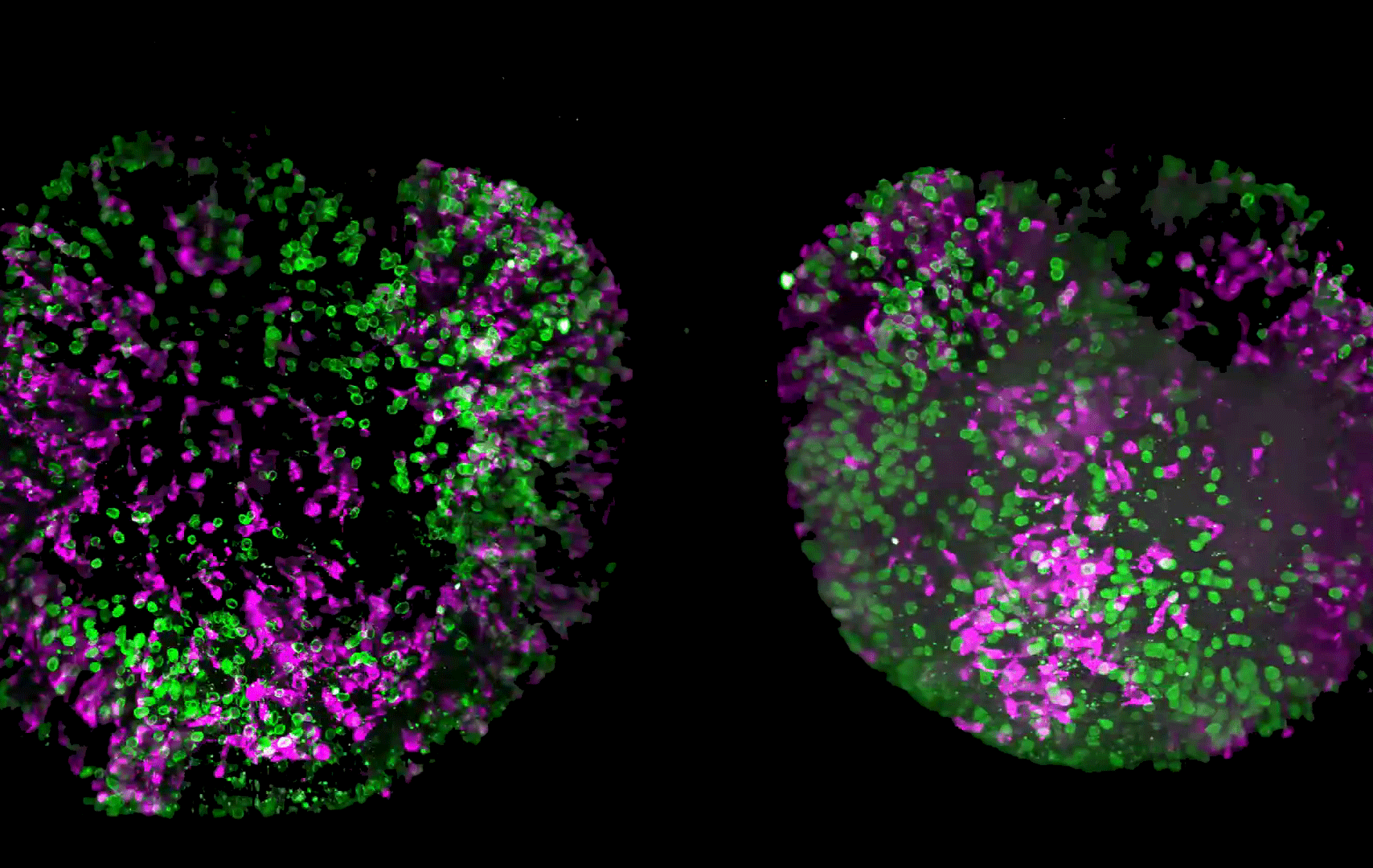
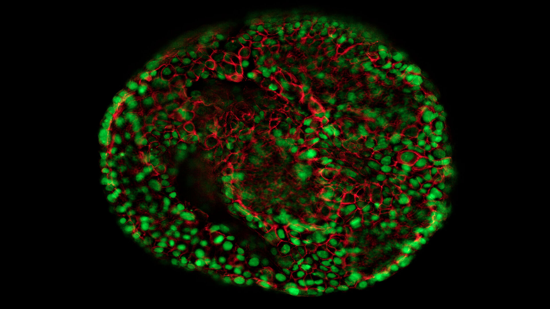
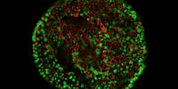
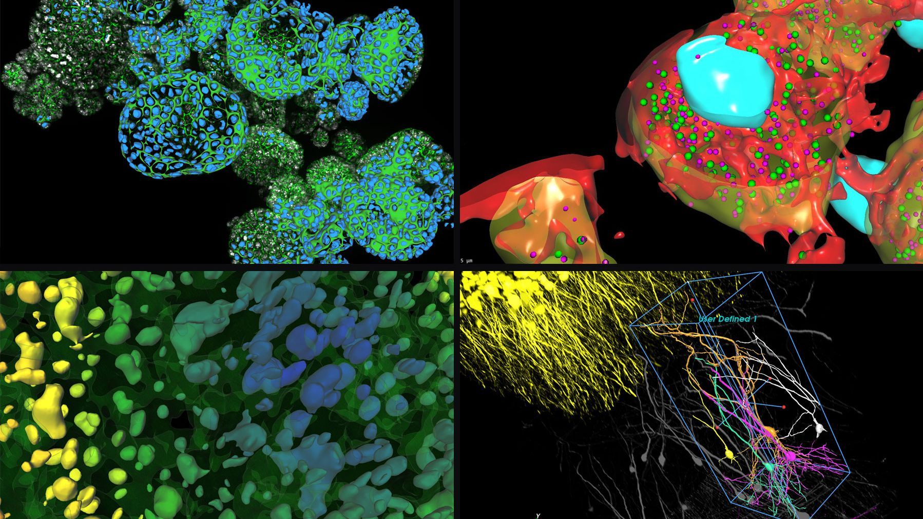
and astrocytes (green) in a cortical spheroid derived from human induced pluripotent stem cells. THUNDER Model Organism Imager with a 2x 0.15 NA objective at 3.4x zoom was used to produce this 425 µm Z-stack (26 positions), which is presented here as an Extended Depth of Field (EDoF) projection.  Images courtesy of Dr. Fikri Birey  from the Dr. Sergiu Pasca laboratory at Stanford University, 3165 Porter Dr., Palo Alto, CA

Download The Guide to Live Cell Imaging

In life science research, live cell imaging is an indispensable tool to visualize cells in a state as in vivo as possible. This E-book reviews a wide range of important considerations to take to…
Primary leaves of cowpea (Vigna unguiculata "California Blackeye") 
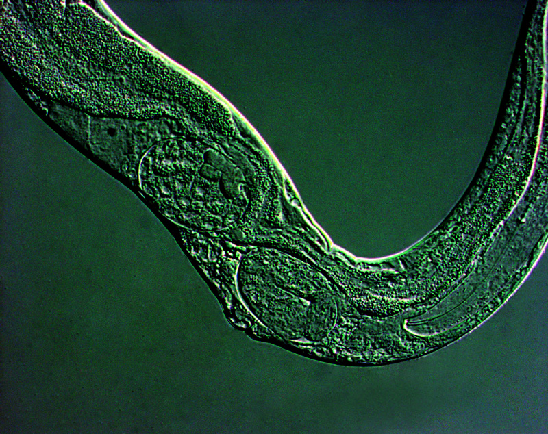
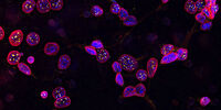
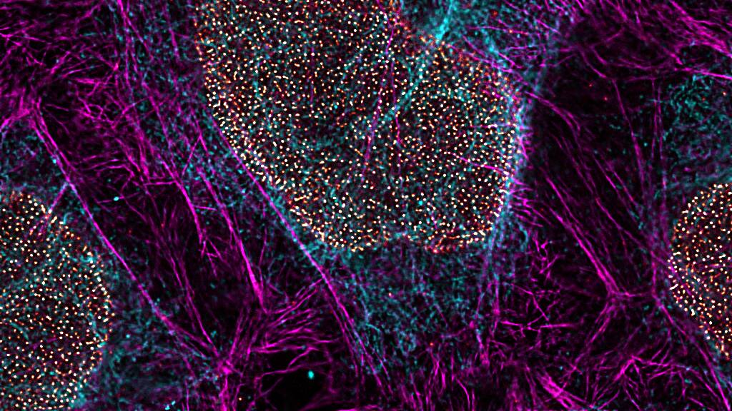
inoculated with cowpea mosaic virus (CPMV) containing the GFP-gene inserted between the movement protein (MP) and the capsid proteins (CPs) in the viral RNA 2

Introduction to Live-Cell Imaging

The understanding of complex and fast cellular dynamics is an important step to get insight into biological processes. Therefore, today’s life science research more and more demands studying…
Image of C2C12 cells: The cells are stained with lamin B (magenta) which indicates nuclear structure, Hoechst (blue) indicating DNA, and γH2AX (yellow) indicating damage to DNA. Cells were imaged using a THUNDER Imager 3D Live Cell with a 63X/1.4 oil immersion objective.

Cell Biology Research

Choosing the right microscopy method is critical if you want to maximize your research output and obtain the highest quality data. Leica Microsystems offers various solutions to improve your cell biology research.

Read More

Introducing Coral Cryo workflow

Correlative Light and Electron Microscopy Solutions

Correlation of Light and Electron Microscopy is crucial for a successful experiment, both for room temperature and cryo-CLEM experiments. CLEM solutions from Leica Microsystems ensure sample viability and quality checks.

Read More

Cell Culture

When it comes to cell and tissue cultures, there are some important differences between the solutions. Leica cell-and-tissue-culture microscopes offer you the ease of use and flexibility with contrast methods that you need for your individual requirements.

Read More

Organoids and 3D Cell Culture

From traditional methods, such as widefield or confocal microscopy, to more advanced imaging methods, such as multiphoton imaging or LightSheet, Leica Microsystems makes it possible to visualize fine cellular details as well as overall tissue architecture within your 3D cell cultures.

Read More

Image of C2C12 cells: The cells are stained with lamin B (magenta) which indicates nuclear structure, Hoechst (blue) indicating DNA, and γH2AX (yellow) indicating damage to DNA. Cells were imaged using a THUNDER Imager 3D Live Cell with a 63X/1.4 oil immersion objective.
Introducing Coral Cryo workflow
Scroll to top