ATTO-TEC Consumables

ATTO-TEC Consumables

ATTO-TEC Consumables

ATTO dyes have become a benchmark for fluorescence microscopy imaging, offering a highly differentiated panel. Their brightness and photostability make them the reagents of choice for demanding applications.

About ATTO-TEC

ATTO-TEC GmbH was founded in autumn 1999 as a spin-off of the University of Siegen. The decisive factor was the scientific experience of the four founders with organic laser and fluorescent dyes, some of which lasted decades. 

In particular, Prof. Drexhage's research team focused on the synthesis and development of long-wavelength fluorophores for the labeling of biomolecules in the 1990s, as fluorescence-based techniques were established as sensitive detection and analysis methods in the emerging field of life sciences. Thus, in the summer of 2000, the young company was able to present its own market-ready products as a manufacturer of fluorescent dyes for medicine, diagnostics and biology at ACHEMA in Frankfurt am Main.

ATTO-TEC was acquired by Leica in 2024 and is a wholly owned subsidiary of Leica Microsystems.

Contact Us

Contact a local imaging specialist for expert advice on the right consumables for your needs and budget.

Key Benefits

High Photostability and Brightness

ATTO dyes are known for their superior photostability and brightness, making them ideal for biological imaging and analysis.

Versatility

These dyes are widely used in molecular biology techniques, such as real-time PCR, DNA sequencing, and fluorescence in situ hybridization (FISH).

Unique Molecular Structure

Their rigid chromophore structure ensures consistent optical properties, reducing variations caused by solvents or temperature.

Portfolio of 43 fluorophores with over 400 different modifications

ATTO-TEC GmbH now has a portfolio of 43 fluorophores with over 400 different modifications. These partially patented fluorescence markers cover the entire visible spectral range from ultraviolet to near infrared.

Our customers – including renowned research institutes and international companies – appreciate the particularly high purity and excellent quality of our ATTO dyes. In addition to our readily available catalogue products, we offer the service of individual customer syntheses. In close cooperation with the client, our qualified team develops tailor-made dyes or derivatives for its special application.

The satisfaction of our customers is our top priority. Please do not hesitate to contact us if you have any problems or questions regarding our particular products as well as fluorescence or organic dyes. We will be glad to help you!

Related Articles

Read our latest articles about Fluorescence

The knowledge portal of Leica Microsystems offers scientific research and teaching material on the subjects of microscopy. The content is designed to support beginners, experienced practitioners and scientists alike in their everyday work and experiments.

More Articles
Mouse fibroblasts where F-actin is labelled with FITC (green)
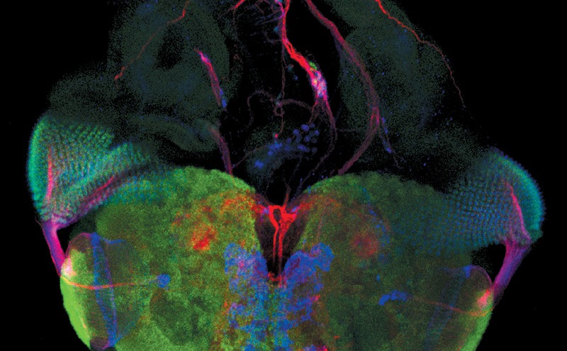
, tubulin with Cy5 (red), and nuclei with DAPI (blue). Image courtesy of Dr. Günter Giese, Max Planck Institute for Medical Research, Heidelberg, Germany.

Overview of Fluorescent Dyes in terms of Applications and Properties

An introduction to commonly used fluorescent dyes and an overview of their characteristics are given in this article. Fluorescence microscopy is used for the study of specific cellular components with…
Zebrafish heart, DAPI (nuclei, blue), Tropomyosin (cardiomyocytes, red) and GFP (primordial cardiac layer, green). Courtesy of Anna Jazwinska, University of Fribourg, Switzerland.

A Guide to Fluorescence Microscopy

Fluorescence microscopy uses the ability of fluorophores, dyes, or fluorescent proteins to emit light of a specific wavelength after being excited with light of a shorter wavelength. Biomolecules can…
Optical microscope image, which is a composition of both brightfield and fluorescence illumination, showing organic contamination on a wafer surface. The inset images in the upper left corner show the brightfield image (above) and fluorescence image (below with dark background).

Visualizing Photoresist Residue and Organic Contamination on Wafers

As the scale of integrated circuits (ICs) on semiconductors passes below 10 nm, efficient detection of organic contamination, like photoresist residue, and defects during wafer inspection is becoming…
Plastic reconstructive surgery with M530 OHX

Advances in Oncological Reconstructive Surgery

Decision making and patient care in oncological reconstructive surgery have considerably evolved in recent years. New surgical assistance technologies are helping surgeons push the boundaries of what…
Brain organoid labeled with lamin (green) and tubulin (magenta), acquired using Viventis Deep. Courtesy of Akanksha Jain, Treutlein Lab ETH-DBSSE Basel (Switzerland).

Faster & Deeper Insights into Organoid and Spheroid Models

Gain deeper, more translatable, insights into organoid and spheroid models for drug discovery and disease research by overcoming key imaging challenges. In this eBook, explore advanced microscopy…
Image of roundworm C. elegans acquired with a M205 FA fluorescence automated stereo microscope in combination with Rottermann contrast. Areas labelled with mCherry are seen as reddish purple.

A Guide to C. elegans Research – Working with Nematodes

Efficient microscopy techniques for C. elegans research are outlined in this guide. As a widely used model organism with about 70% gene homology to humans, the nematode Caenorhabditis elegans (also…

A Novel Laser-Based Method for Studying Optic Nerve Regeneration

Optic nerve regeneration is a major challenge in neurobiology due to the limited self-repair capacity of the mammalian central nervous system (CNS) and the inconsistency of traditional injury models.…
Fluorescence microscopy of sectioned tissue, showing the interface between the extensor digitorum longus muscle and the common peroneal nerve in the adult rat. Regenerative peripheral nerve interface (RPNI) at 2 weeks. Image acquired using Mica. Stained for nuclei (blue), neurofilaments (green) and S100B (red). Image courtesy of Dr. Aaron Lee, Department of Bioengineering (Lab of Dr. Rylie Green), Imperial College London.

How to Image Axon Regeneration in Deep Muscle Tissue

This study highlights Dr. Aaron Lee’s research on mapping nerve regeneration in muscle grafts post-amputation. Limb loss often leads to reduced quality of life, not only from tissue loss but also due…
Scroll to top