Automated Microscopes

Automated Microscopes

Automated Microscopes

Automated microscopes are very useful for applications which require many repetitious observations over a long period of time. Examples are live-cell imaging or high-throughput analysis for quality assurance. Leica automated microscopes help life scientists and quality engineers increase the efficiency of their workflows.

Read our latest articles about Automated Microscopes

The knowledge portal of Leica Microsystems offers scientific research and teaching material on the subjects of microscopy. The content is designed to support beginners, experienced practitioners and scientists alike in their everyday work and experiments.

More Articles

Mica: A Game-changer for Collaborative Research at Imperial College London

This interview highlights the transformative impact of Mica at Imperial College London. Scientists explain how Mica has been a game-changer, expanding research possibilities and facilitating interdisciplinary collaboration. They explain how detailed live cell imaging with Mica provides more meaningful information, keeping scientists at the forefront of research. The team foresees Mica continuing to open new research avenues, including the study of microfluidics and other advanced applications.

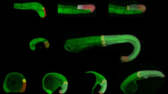
How to Study Gene Regulatory Networks in Embryonic Development

Join Dr. Andrea Boni by attending this on-demand webinar to explore how light-sheet microscopy revolutionizes developmental biology. This advanced imaging technique allows for high-speed, volumetric live imaging of 3D samples with minimal phototoxicity. Learn through user examples how light-sheet microscopy is enhancing our understanding of intestinal and brain organoid development and dive into the technology behind the Viventis Deep microscope from Leica Microsystems and its application in long-term imaging.

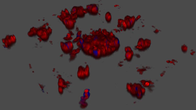
Cutting-Edge Imaging Techniques for GPCR Signaling

With this webinar on-demand enhance your pharmacological research with our webinar on GPCR signaling and explore cutting-edge imaging techniques that aim to understand how GPCR signaling translates into cellular and physiological responses. Discover leading research that's expanding what we know about these critical pathways to find new avenues for drug discovery.

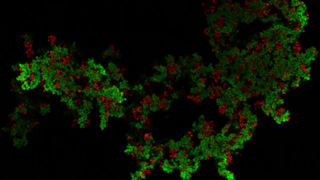
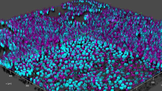
Exploring Microbial Worlds: Spatial Interactions in 3D Food Matrices

The Micalis Institute is a joint research unit in collaboration with INRAE, AgroParisTech, and Université Paris-Saclay. Its mission is to develop innovative research in the field of food microbiology for health. In this video series, researchers from the Micalis Institute explain how Mica is used in various microscopy experiments to investigate the interactions between food, foodborne microorganisms, and the host. Using Mica,enables rapid, high-resolution 3D imaging of food matrices and microbial interactions, they can better understand the emerging properties of these spatially organized communities.

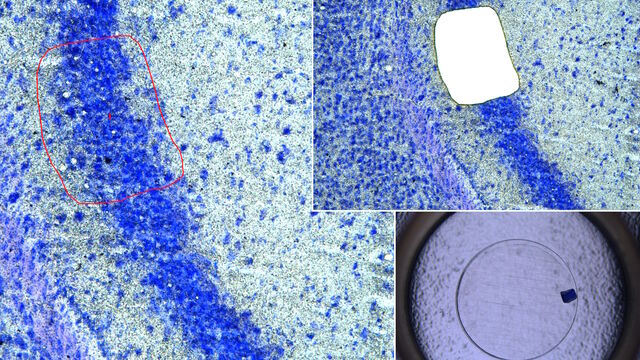
Molecular Biology Analysis facilitated with Laser Microdissection (LMD)

Extracting biomolecules, proteins, nucleic acids, lipids, and chromosomes, as well as extracting and manipulating cells and tissues with laser microdissection (LMD) enables insights to be gained into the function of genes and proteins. It is used for a range of applications in neurobiology, immunology, developmental, cell biology, and forensics, e.g., research on cancer and diseases, genetic modifications, molecular pathology and biology, and biochemistry. LMD is also useful for studying protein functions, molecular mechanisms, and their interaction in transduction pathways.

Rapidly Visualizing Magnetic Domains in Steel with Kerr Microscopy

The rotation of polarized light after interaction with magnetic domains in a material, known as the Kerr effect, enables the investigation of magnetized samples with Kerr microscopy. It allows rapid visualization of magnetic domains at the material’s surface. For efficient R&D and QC of magnetic materials, e.g., steel alloys, used in electrical and electronic devices, Kerr microscopy can play an important role. More details about how Kerr microscopy can be used to image magnetic domains in the grains of steel alloys is described in this article.

Leveraging AI for Efficient Analysis of Cell Transfection

This article explores the pivotal role of artificial intelligence (AI) in optimizing transfection efficiency measurements within the context of 2D cell culture studies. Precise and reliable transfection efficiency measurements for 2D cell culture are key for understanding cellular mechanisms. A high transfection efficiency of the targeted proteins is crucial for experiments including live-cell imaging and protein purification. Manual estimation is inconsistent and unreliable. With the power of AI, efficient and reliable transfection studies can be achieved.

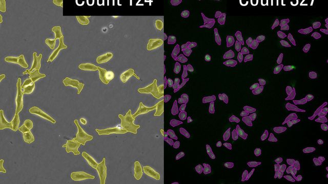
Precision and Efficiency with AI-Enhanced Cell Counting

This article describes the use of artificial intelligence (AI) for precise and efficient cell counting. Accurate cell counting is important for research with 2D cell cultures, e.g., cellular dynamics, drug discovery, and disease modelling. Precise cell counting is critical for determining cell viability, proliferation rates, and the effects of experimental conditions. These factors are essential for reliable and robust results. How an AI-based approach can significantly enhance the accuracy and speed of cell counting, leading to a significant impact on cellular research, is described.

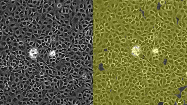
AI Confluency Analysis for Enhanced Precision in 2D Cell Culture

This article explains how efficient, precise confluency assessment of 2D cell culture can be done with artificial intelligence (AI). Assessing confluency, the percentage of surface area covered, accurately is crucial for reliable cell research. Traditional methods use visual inspection or simplistic algorithms, making precision challenging, especially with complex cell lines used for drug discovery, tissue engineering, and regenerative medicine. Methods exploiting AI with automated image analysis and deep-learning algorithms offer better precision and can enhance experimental results.

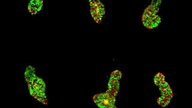
Dual-View LightSheet Microscope for Large Multicellular Systems

Visualizing the dynamics of complex multicellular systems is a fundamental goal in biology. To address the challenges of live imaging over large spatiotemporal scales, Franziska Moos et. al. present an open-top multisample dual-view lightsheet microscope in a paper published in Nature Methods. The authors find that the Viventis Deep microscope offers significant advances in imaging large samples with single-cell resolution.

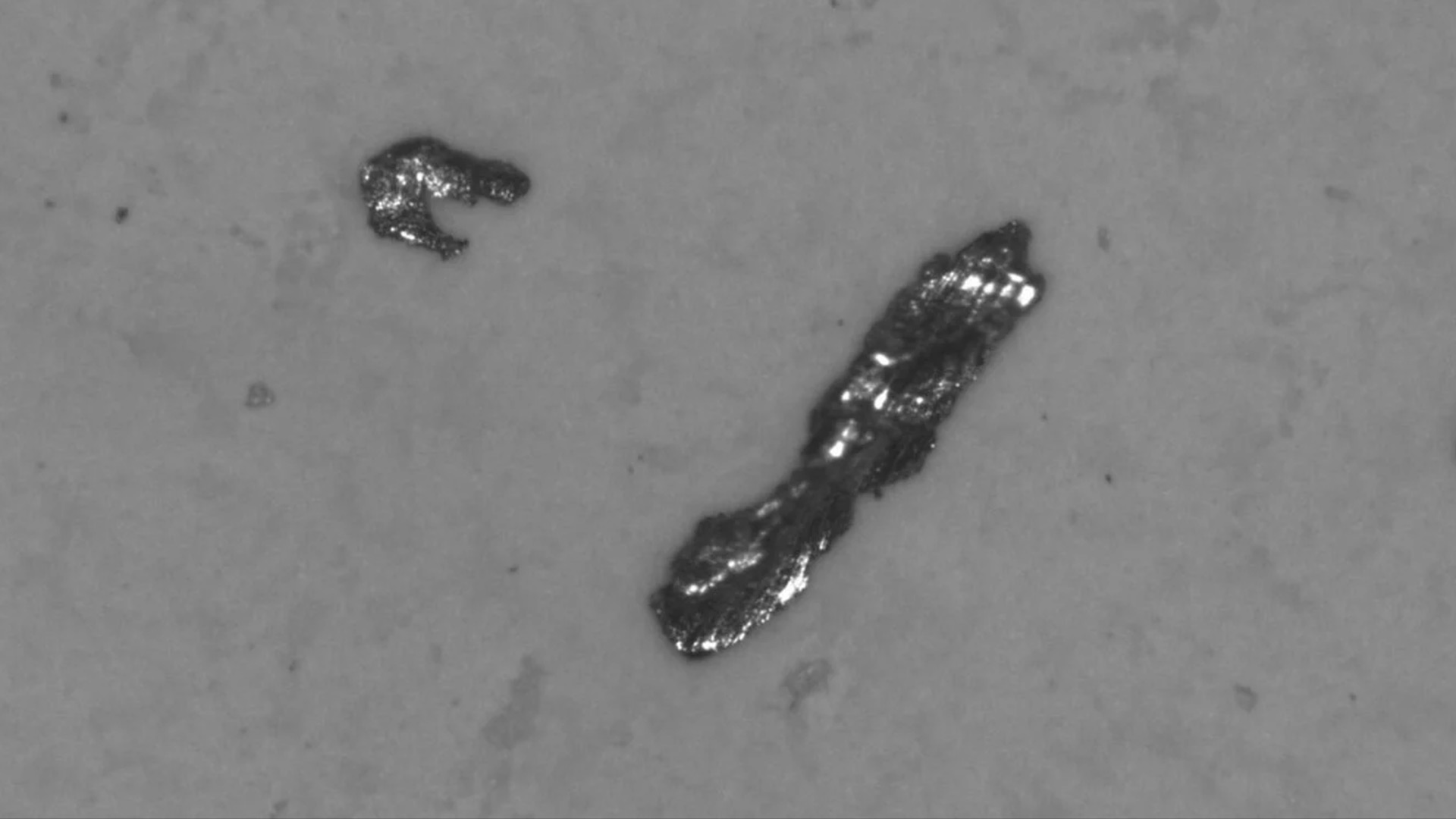
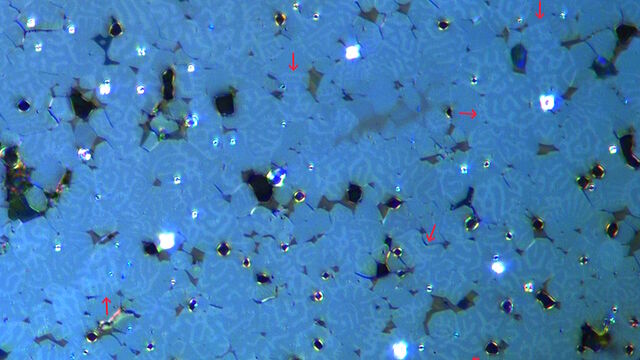
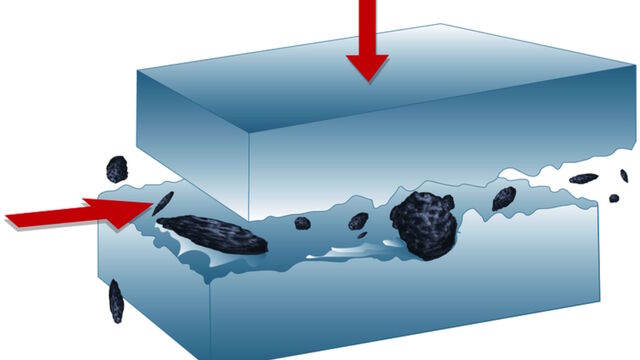
Battery Particle Detection During the Production Process

How battery particle detection and analysis is enhanced with optical microscopy and laser spectroscopy for rapid, reliable, and cost-effective QC during battery production is explained in this article.

Rapid Check of Live Stem Cells in Cell-Culture Inserts set in Multi-Well Plates

See how efficient imaging of live iPSC stem cells within cell-culture inserts set in a multi-well plate can be done to evaluate the cells using a THUNDER Imager. Just read this article.

How to Prepare Samples for Stimulated Raman Scattering (SRS) imaging

Find here guidelines for how to prepare samples for stimulated Raman scattering (SRS), acquire images, analyze data, and develop suitable workflows. SRS spectroscopic imaging is also known as SRS microscopy.

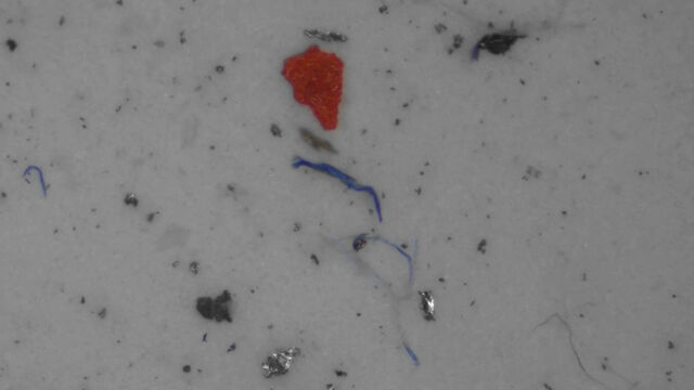
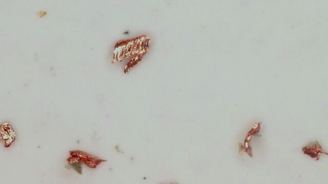
Key Factors for Efficient Cleanliness Analysis

An overview of the key factors necessary for technical cleanliness and efficient cleanliness analysis concerning automotive and electronics manufacturing and production is provided in this article.

Notable AI-based Solutions for Phenotypic Drug Screening

Learn about notable optical microscope solutions for phenotypic drug screening using 3D-cell culture, both planning and execution, from this free, on-demand webinar.

Five Inverted-Microscope Advantages for Industrial Applications

With inverted microscopes, you look at samples from below since their optics are placed under the sample, with upright microscopes you look at samples from above. Traditionally, inverted microscopes are used for life science research, because gravity makes samples sink to the bottom of a holder with aqueous solution and you don’t see a lot from above.

Key Questions

1What is an automated microscope?

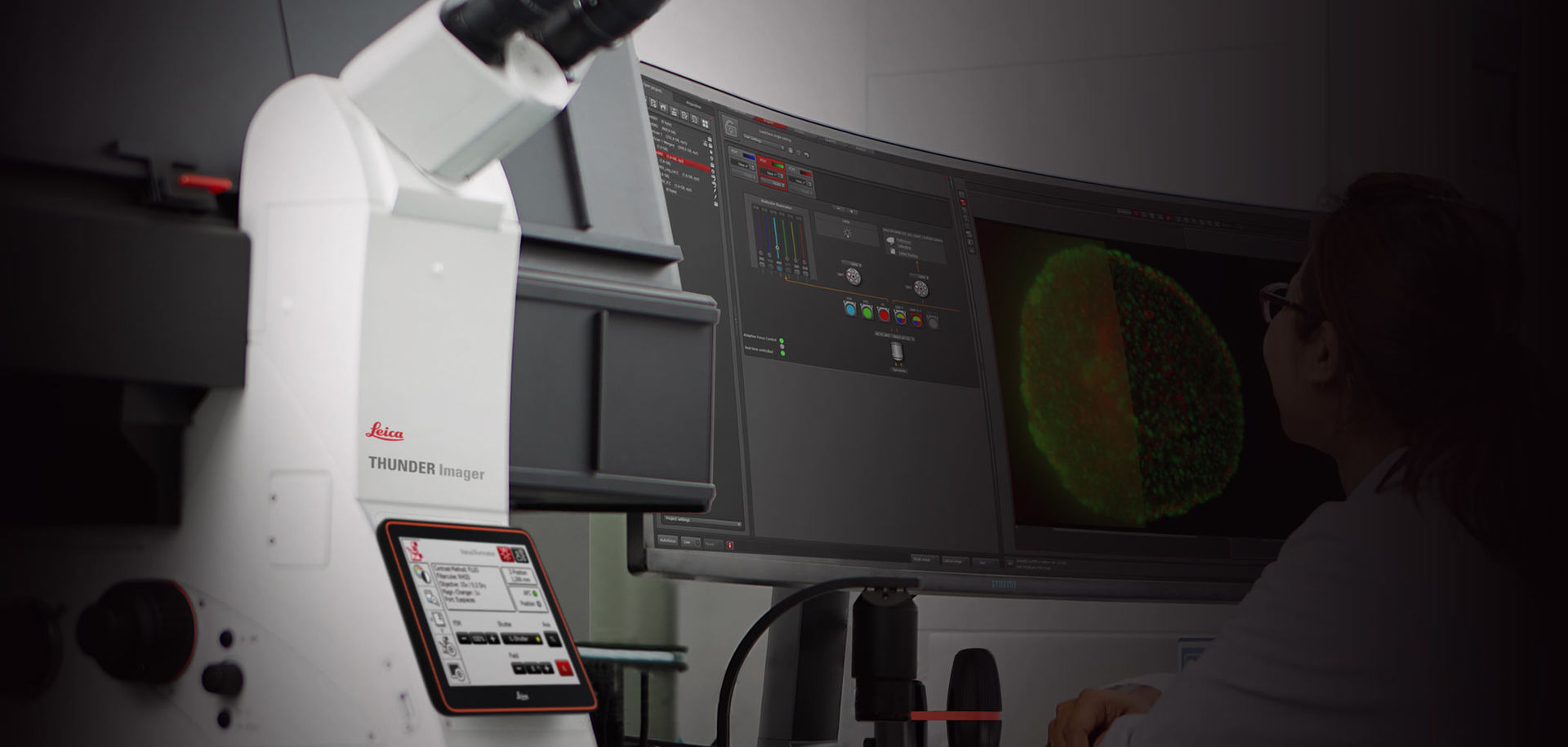
An automated microscope automates repetitious and time-consuming tasks, thereby enabling a higher throughput and faster acquisition of accurate and reliable images. It also helps users save time, because there is much less need of intervention. The microscope uses electronic components, like a digital camera, and advanced software to perform automatically operations, like sample movement, objective changing, autofocus, illumination, etc.

2What is automated imaging?

Automated imaging is a more efficient way to acquire images using an automated microscope. It enables high-speed and time-lapse imaging using advanced softwares without the requirement of manual intervention. To save time during imaging, the microscope automates several common tasks, e.g., focusing, sample movement, changing illumination settings, changing objectives, etc., to minimize or eliminate laborious and time-consuming user operations.

3What is a smart microscope?

A smart microscope can perform several tasks automatically without the need of user intervention. Examples are autofocusing, illumination settings, sample movement, changing objectives, etc. It uses electronic components, e.g., a digital camera, motorized stage, and computer interfaces, and advanced software in order to provide faster acquisition of accurate and reliable images.

Automated microscope DMi 8

Why automated microscopes?

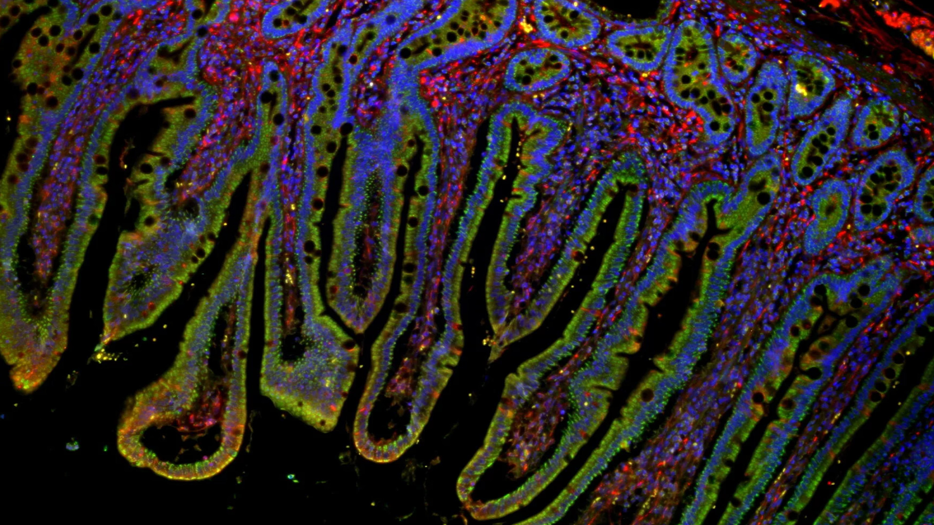
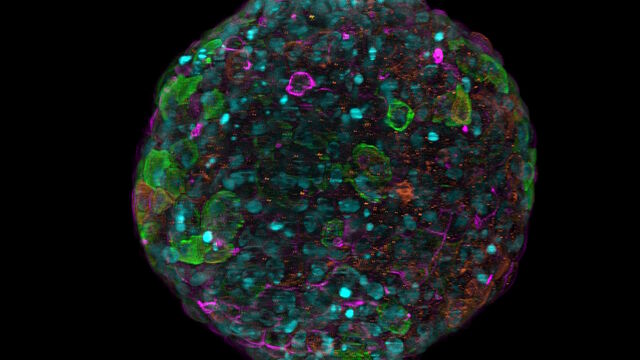
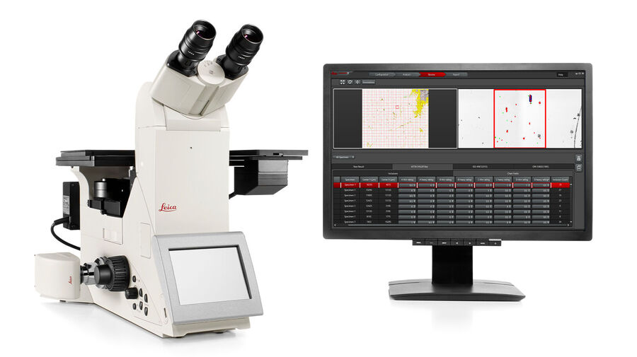
Automated microscopes are useful for life science fluorescence applications like live-cell and time-lapse imaging (widefield and confocal microscopy) as well as high-speed multi-fluorescence optical sectioning (confocal microscopy). They are also practical for industrial applications like cleanliness analysis, alloy quality rating, and optical inspection of electronic circuit boards (compound, digital, and stereo microscopes). The microscopes have features, such as automated contrast and illumination including fluorescence excitation and emission, motorized focus and parfocality, automatic brightness and diaphragm adjustment, among others. These automated functions make the microscopes more convenient to use and lead to more reliable and reproducible results.

The advantages of using automated microscopes

Automated microscopes offer advantages over manually operated microscopes for certain applications, as repetitious, labor-intensive, time-consuming operations are automated and do not require user intervention. Automated microscopes also enable high-speed operations to be performed more easily and more reliably. They save a considerable amount of time compared to manual operation and enable a faster and more reliable workflow.

No wasted time: electronic components, digital camera, and intelligent software

An automated microscope requires automated functions

An automated microscope actually has certain functions which are automated. The automation is done by using electronic components, like a digital camera, and intelligent software. For example, focusing, illumination settings, sample movement, changing objectives, can all be automated. The image acquisition time is synchronized with exposure of the sample to light to minimize photodamage if the sample is light-sensitive.

Applications for automated microscopes

Automated microscopes are useful for various applications that require time-consuming and repetitive tasks or high-speed operations. Examples are time-lapse imaging with fluorescence microscopy for life-science applications and inspection and quality control for industrial applications.
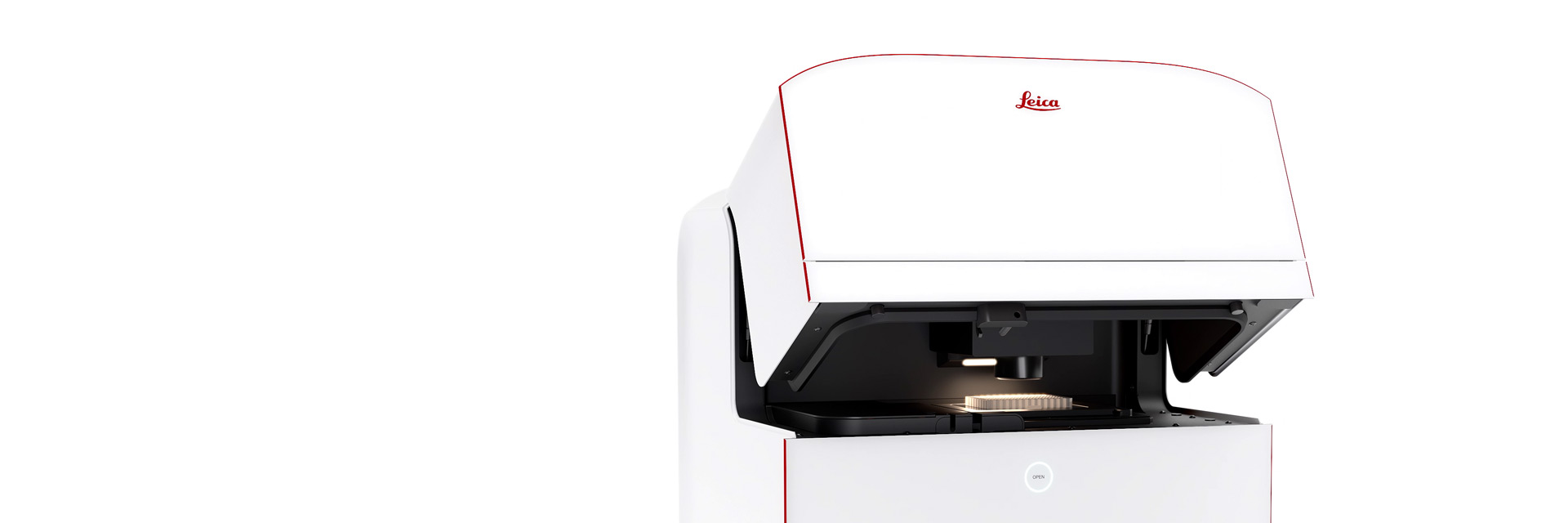
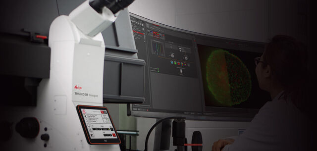
Meet Mica
The world’s first Microhub

Mica enables microscopy access for all, removes the constraints of traditional four-color fluorescence imaging and radically simplifies workflows.

Read more

Frequently Asked Questions Automated Microscopes

When choosing an automated microscope, one should determine which tasks or operations in the workflow need to be automated in order to save time and increase accuracy and reproducibility. Once the tasks are known, then a microscope with the appropriate automated features, e.g., autofocusing, motorized stage for sample movement, control of illumination settings, changing objectives, etc., can be chosen.

A manual microscope uses optical, electronic, and mechanical components (stage, focusing, illumination, etc.) that need to be operated manually by the user. In an automated microscope, these tasks are automated and do not require the operator’s presence during imaging. An automated microscope can provide more accurate and reliable results and lead to a more efficient and less labor-intensive workflow for users.

Automated microscopes use a combination of electronic components and software which enable automation of repetitious tasks being done in the imaging workflow. Common electronic components for microscope automation are digital cameras, motorized stages for sample movement and focusing, motorized objective nosepieces, automated control of illumination settings, etc.

Automated microscopes are useful for imaging tasks which are repetitious and time-consuming when done manually. Performing such tasks with an automated microscope saves users time and enhances reproducibility of the results. Automation is often needed for life-science applications requiring time-lapse imaging over multiple samples, but also for cleanliness inspection and quality control for electronic components.

Technical Cleanliness Analysis Steel Quality

Cleanliness analysis

Cleanliness analysis is key for assuring product quality and performance for automotive and electronic components. Automated microscopes are critical for efficient and reliable cleanliness analysis and the characterization of particulate contamination.

Read More

 Mica widefield image of intestine tissue specimen at 10x magnification. Nuclei are labeled blue, mitochondria green, and detyrosinated tubulin red.

Live-cell imaging

Automated microscopes are very useful for the imaging, analysis, and documentation of live cell and tissue cultures. They help life scientists save time and effort when the research requires repetitive, labor-intensive imaging of multiple live-cell specimens.

Read More

 Mica widefield image of intestine tissue specimen at 10x magnification. Nuclei are labeled blue, mitochondria green, and detyrosinated tubulin red.
Technical Cleanliness Analysis Steel Quality
Scroll to top