Visoria P Polarization Microscope
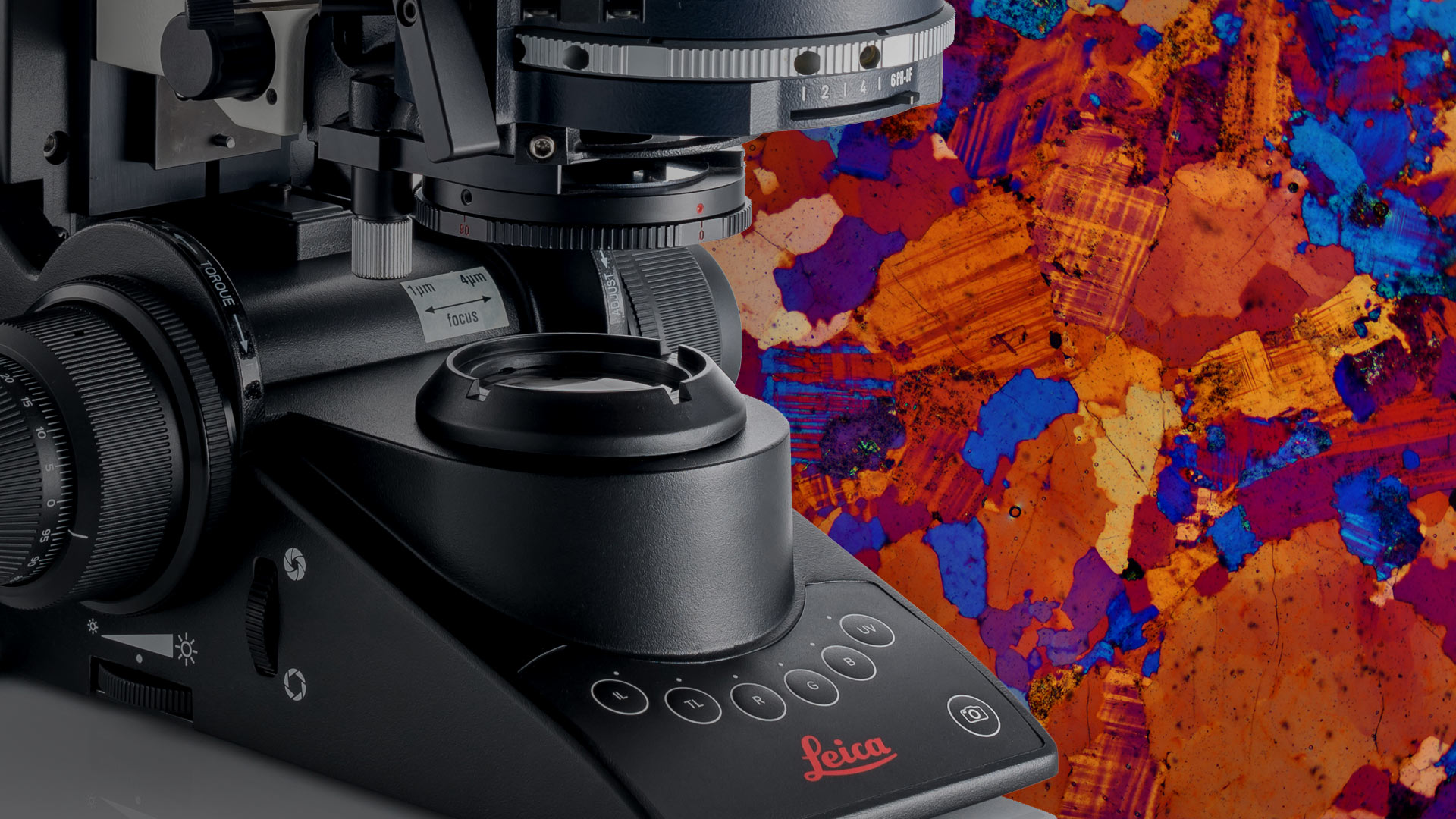
Experience enhanced efficiency and comfort in your daily microscopy routine. The Visoria P polarization microscope uses polarized light to study the optical properties of materials and geological samples.
Streamline your workflows with encoded functions, optimized light settings, and other microscope features. You can also be more comfortable and minimize strain thanks to the microscope’s ergonomic design.
Popular Configurations
")
Visoria P Digital Polarization Microscope for Industrial Applications (Transparent samples only)Experience enhanced efficiency and comfort in your daily microscopy routine. The Visoria P polarization microscope uses polarized light to study the optical properties of materials and geological samples. Streamline your workflows with encoded functions, optimized light settings, and other microscope features. You can also be more comfortable and minimize strain thanks to the microscope’s ergonomic design.
Loading...
View product details+
Product includes
Loading...
|
||||||||||||||||||||||||||||||||||||||||||||||||||||||||||||||||||||||||
Visoria P Polarization Microscope for Geosciences Including Conoscopy with Flexacam c5Experience enhanced efficiency and comfort in your daily microscopy routine. The Visoria P polarization microscope uses polarized light to study the optical properties of materials and geological samples. Streamline your workflows with encoded functions, optimized light settings, and other microscope features. You can also be more comfortable and minimize strain thanks to the microscope’s ergonomic design.
Loading...
View product details+
Product includes
Loading...
|
||||||||||||||||||||||||||||||||||||||||||||||||||||||||||||||||||||||||
Visoria P Polarization Microscope for Industrial Applications with Flexacam c5 (transparent samples only)Experience enhanced efficiency and comfort in your daily microscopy routine. The Visoria P polarization microscope uses polarized light to study the optical properties of materials and geological samples. Streamline your workflows with encoded functions, optimized light settings, and other microscope features. You can also be more comfortable and minimize strain thanks to the microscope’s ergonomic design. This configuration includes the Visoria P polarization microscope as well as Flexacam c5 microscope camera and is suited for industrial applications (transparent samples only)
Loading...
View product details+
Product includes
Loading...
|
||||||||||||||||||||||||||||||||||||||||||||||||||||||||||||||||||||||||

Don’t see your Configuration? Request for individual Quote
Show popular configurations
Explore and buy pre-configured microscopy solutions in our online shop. Enjoy a seamless online shopping experience.
Meet Visoria P
For polarization microscopy
The Visoria P polarization microscope delivers key results when investigating birefringent (optically anisotropic) materials like minerals, rocks, coal, plastics, polymers, liquid crystals, glass,and concrete.
Save time with optimized light settings
Spend more time viewing and examining samples with Visoria P.
If you change the microscope’s magnification or contrast method, there is no need to manually adjust the brightness thanks to the light management function. The illumination settings are automatically applied thanks to the microscope’s encoding.
Simplify your documentation
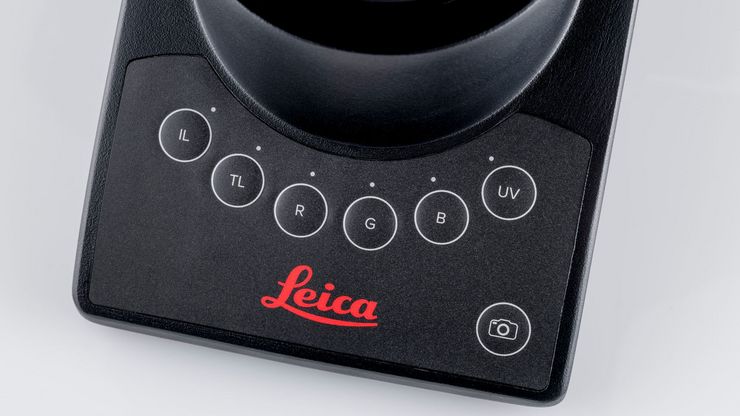
You can quickly capture sample details with a press of a button while keeping your eyes on the image. The button for image acquisition is easily accessible on the Visoria P microscope stand.
When you store an image for documentation, selected system settings are automatically saved along with the meta data of the image.
The scale bar is automatically adjusted and added to the image which increases efficiency and saves you valuable time.
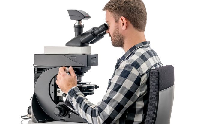
Operate your microscope with ease
Perform daily routines rapidly and reliably thanks to the intuitive operation of Visoria P.
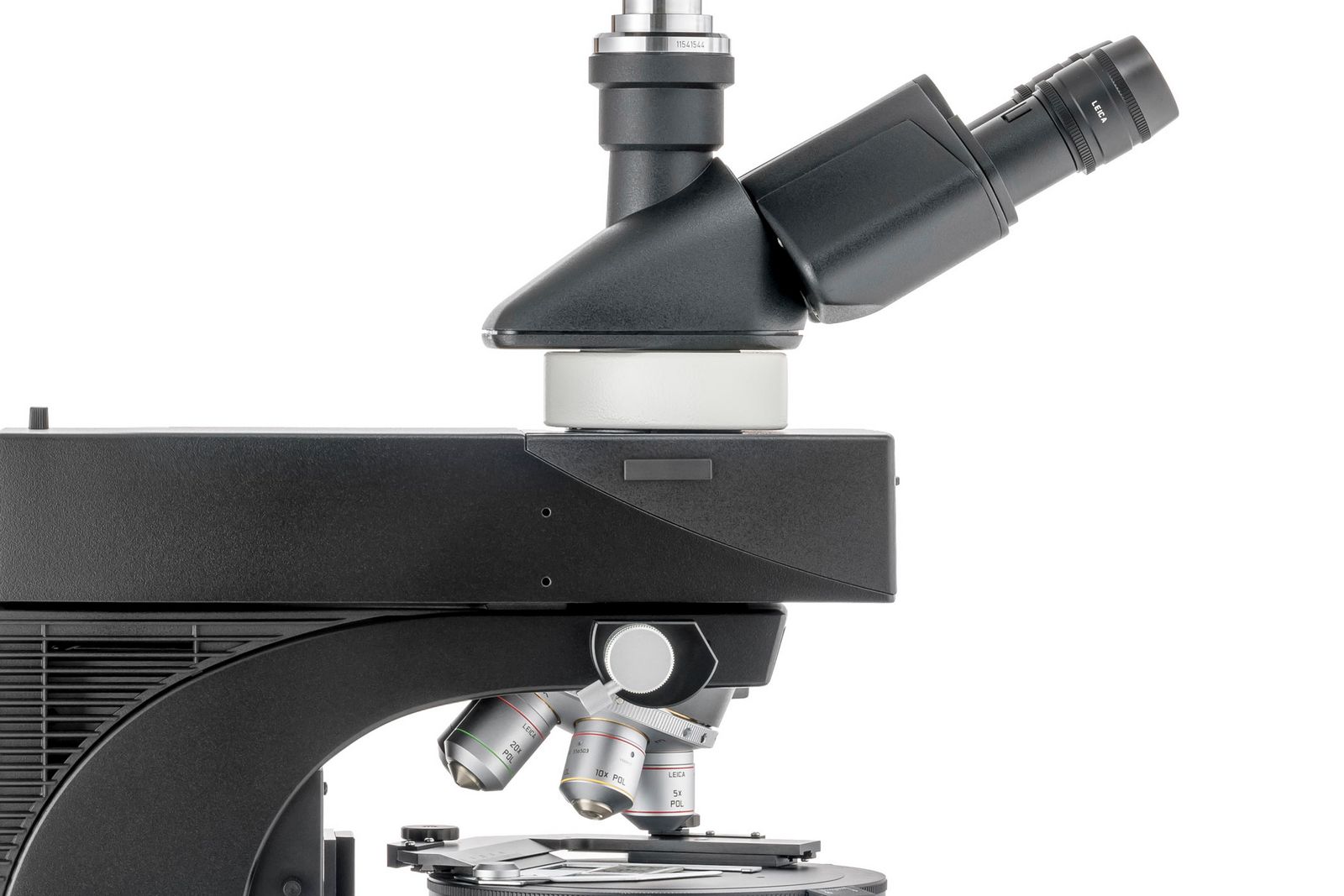
- Easily find the appropriate aperture diaphragm settings for the objective used by matching the color markings.
- Protect your samples and objectives from accidental damage with the built-in focus stop.
- For finer focus at higher magnification, use the three-gear focusing system - coarse, medium, and fine.


The aperture diaphragm's scale on the transmitted-light condenser (left) and incident-light axis (right) has color markings matching the objective color codes.
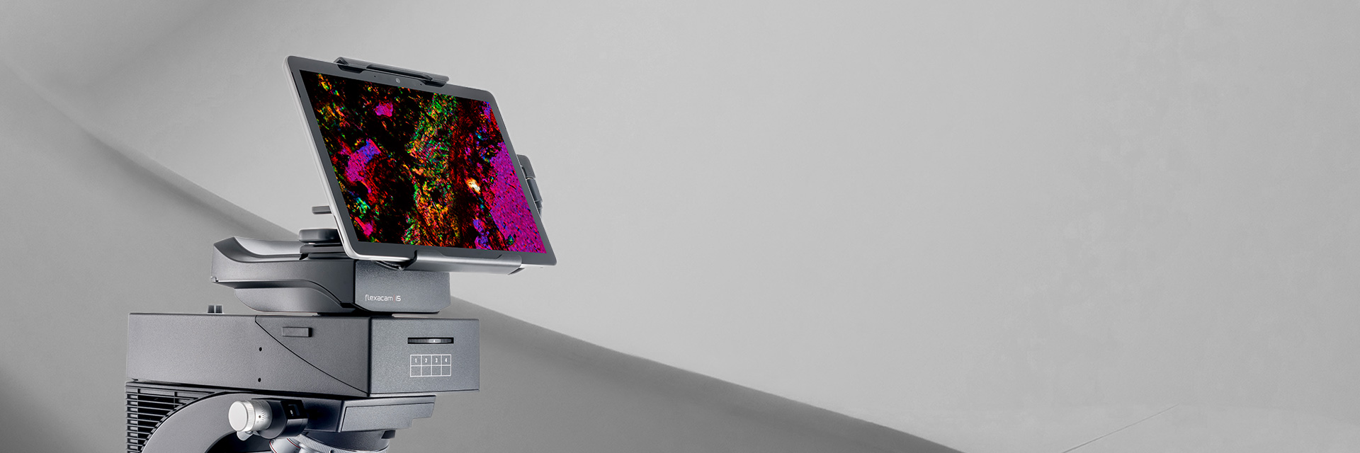
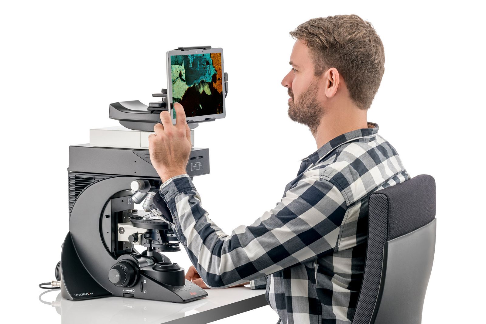
Work without eyepieces by going digital
The Visoria P digital polarization microscope without eyepieces offers a number of practical benefits:
- Work in a comfortable and relaxed position by viewing images directly on a tablet.
- Visualize and document your work steps quickly and discuss image results easily with your colleagues.
- Save space on your workbench without the need for a computer.

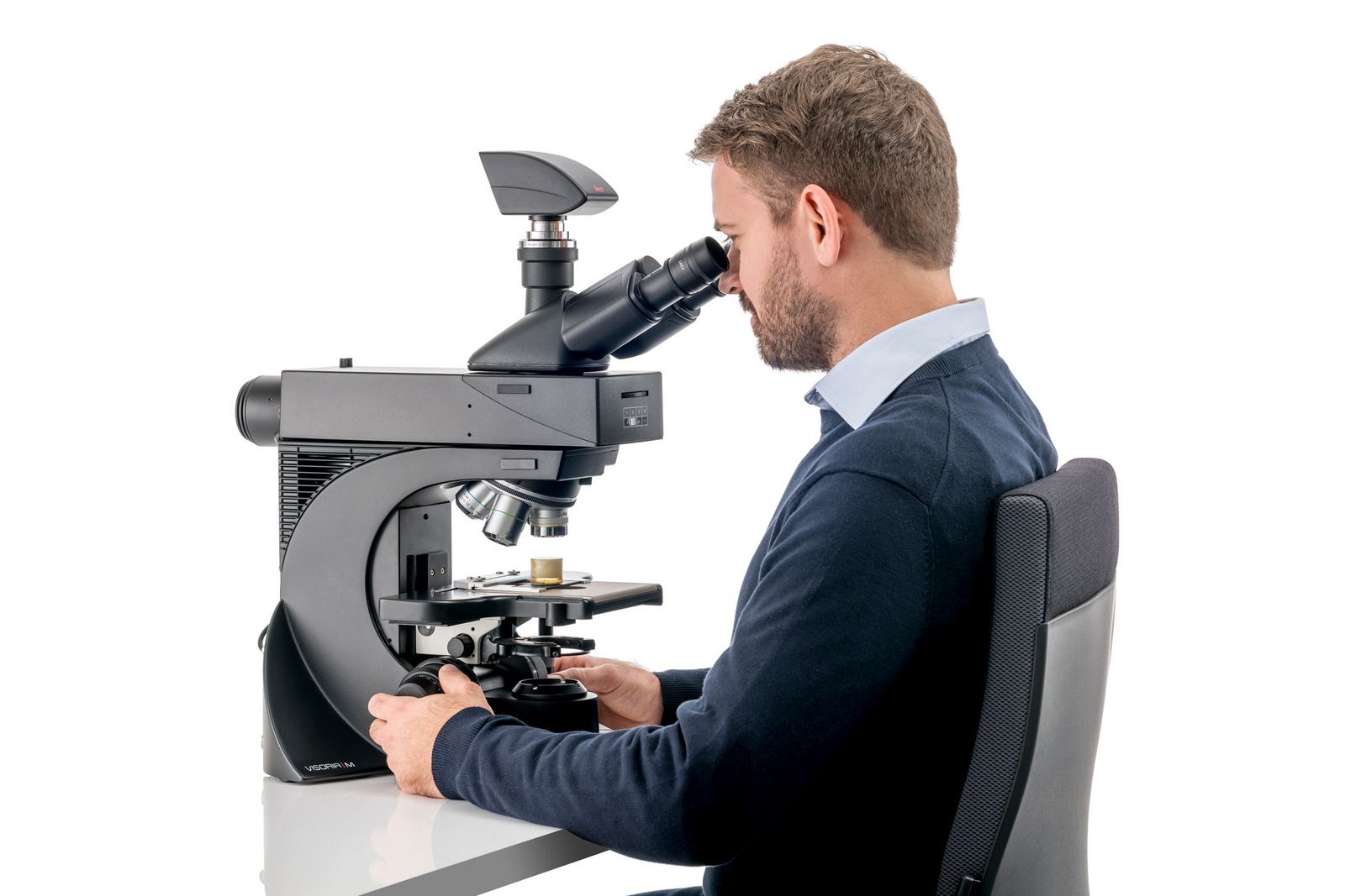
Stay comfortable while working
Visoria P adapts to your needs, allowing a proper posture and reducing the risk of neck and back strain during long hours at the microscope.
Work comfortably with aligned shoulders and optimal hand and arm positioning thanks to the symmetrical layout and height adjustment of the focus control knobs.
Adapt your microscope with ergo accessories
You can maintain an upright posture thanks to the adaptability of Visoria P. Choose from a range of ergonomic accessories to suit your needs.
- Ergonomic modules: Insert ErgoModules below the tube to adjust the eyepiece height for a comfortable sitting posture.
- Ergonomic lift: The optional ErgoLift enables easy height adjustments of the microscope.
Determine optical properties of materials with conoscopy
Conoscopy is useful for evaluating the optical properties of anisotropic materials.
Choose from three conoscopy modules
- Bertrand lens cube
- Bertrand lens module (A/B module)
- Advanced conoscopy module.


Conoscopic image of calcite. Left: Acquired with circular polarized light. The position of the optical axis can be clearly determined with circular polarization. Right: With linear polarized light. The calcite section is perpendicular to the optical axis.
Specialized objectives for coal and asbestos analysis
For the analysis of coal, Visoria P helps you to identify different sample components using specialized oil-immersion objectives.
To determine the presence of asbestos in a sample, the dispersion color method is typically used. For this analysis, you can take advantage of the dispersion staining objectives.


Left: Fibrous actinolite imaged with parallel polarizers. Right: Same actinolite sample imaged with crossed polarizers. The actinolite fibers show prominent colors from birefringence which clearly distinguish them from glass fibers (no birefringence).
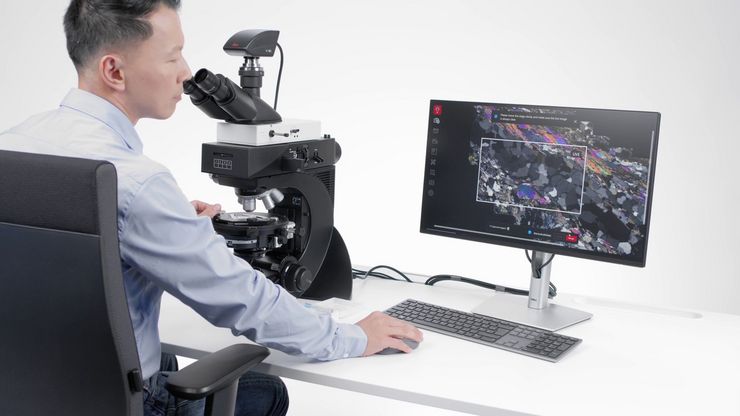

Powered by the Enersight software platform
Simplify and streamline your workflow with the Visoria P polarization microscope and Enersight software platform. It helps you compare, measure, and share seamlessly with a single intuitive interface.
Key advantages
- Observe samples with a larger field of view and higher resolution using the XY Stitching with Manual Stage function.
- Acquire sharp images of samples with extended depth of field (EDOF).
- Capture images with optimal illumination and camera settings by using the Quick Brightness function.
- Optimize images by automatic correction of shading due to uneven illumination.
- Gain a better understanding of samples by merging multiple images from different contrast methods, such as brightfield and polarization.
