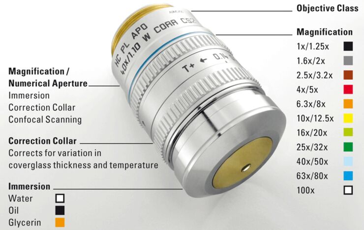
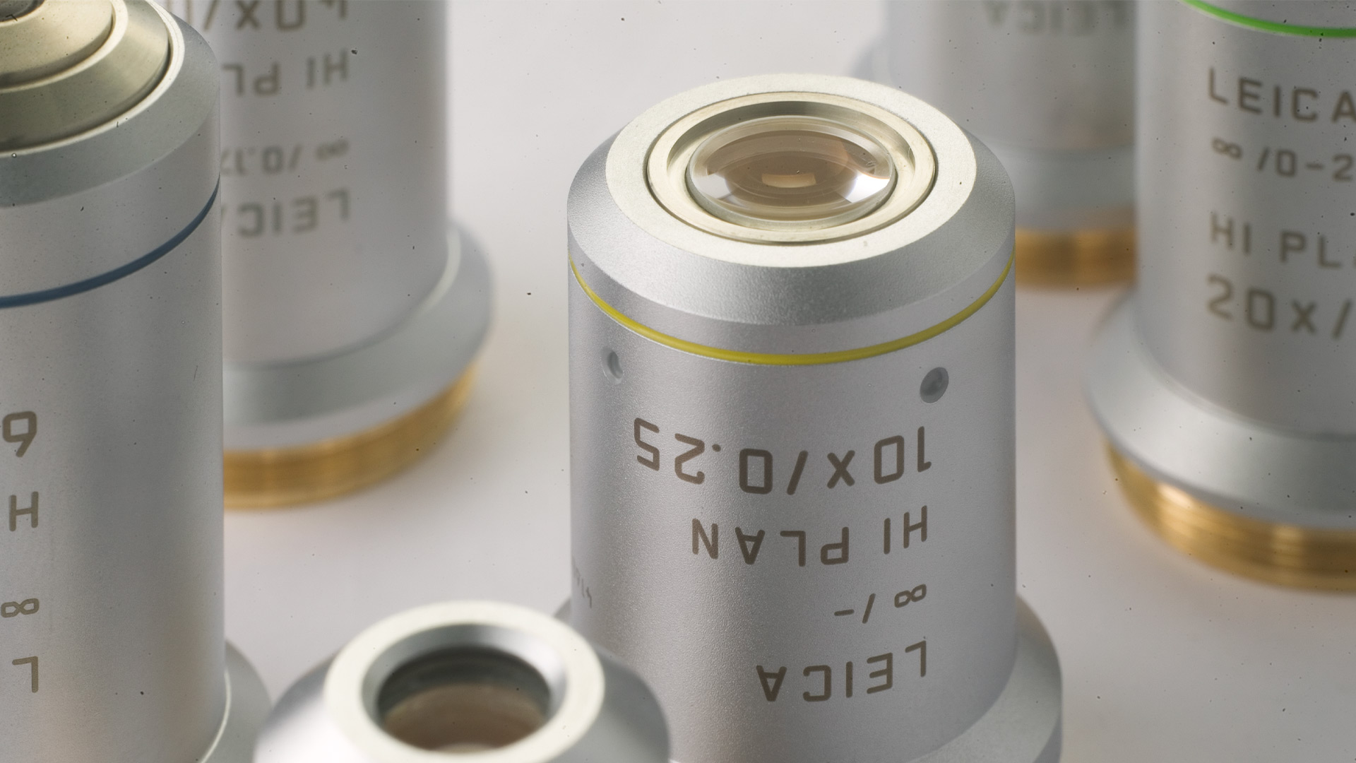
Labeling of Objectives
Leica Microsystems objectives are coded and labeled differently according to type. The coding and labeling provides a short and compact overview for the identification of the objective and for the main optical performances and applications of the objectives.
You can find information about the assignment of the optical systems, e.g. like “HC” and “∞” for infinity corrected optics. Regarding the other indications, please see below in detail.
You have a need for sophisticated, highly specialized and unique lenses? You need small adjustments to existing lenses?
Leica Microsystems HC System
The Leica Microsystems HC system (Harmonic Compound System) comprises the optical components that have been matched to each other for optimal image generation and are involved in the correction of the optical aberrations: objectives, eyepieces, tube lenses, adapter for camera and TV.
For the correction of certain optical aberrations, the microscope is considered as a whole system. Spherical aberrations, coma and axial chromatic aberrations are best corrected at the place where they originate, i.e. in the particular component.
Lateral chromatic aberrations and astigmatism are corrected in parallel in the objective, the tube lens and the eyepiece. The optimal image result is therefore achieved through the interplay of corrections.
HC = The objective is included in the HC system.
HCX = The objective is also compatible with optics of the past (Delta optics 1991-1997)
The HC system ensures
- balanced optical and mechanical fitting dimensions,
- balanced alignment of all optical system components,
- balanced, reliable technical solutions,
- superlative optical performance with progressive manufacturing technology.
Magnification of the Objective
Each objective is labeled with its magnification, for instance 5x or 100x. However, the magnification of the objective alone does not determine the overall magnification of the microscope. This results from the objective magnification multiplied by the eyepiece magnification (for tube lens 1x).
Example:
40x objective x 10x eyepieces = 400x overall magnification
It should be noted, however, that the higher the objective magnification, the lower the visible object field.

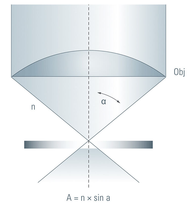
Numerical Aperture
The numerical aperture (NA or A) of the objective is a key parameter for the optical image and determines the resolving power of the objective and the brightness of the image. It is defined by the sine of the half aperture angle a of the lens and the refractive index n of the immersion medium. According to this definition, the larger the numerical aperture, the more narrow the focal spot and hence the higher the resolving power.
The objectives are labeled with their magnification, followed by the particular NA value, for instance 10x/0.40 or 63x/1.40. The numerical aperture of the objective can be changed by using iris diaphragm objectives.
The term 'numerical aperture’ is explained in detail in the Leica Science Lab article: "Beware of "Empty" Magnification"
Iris Diaphragm
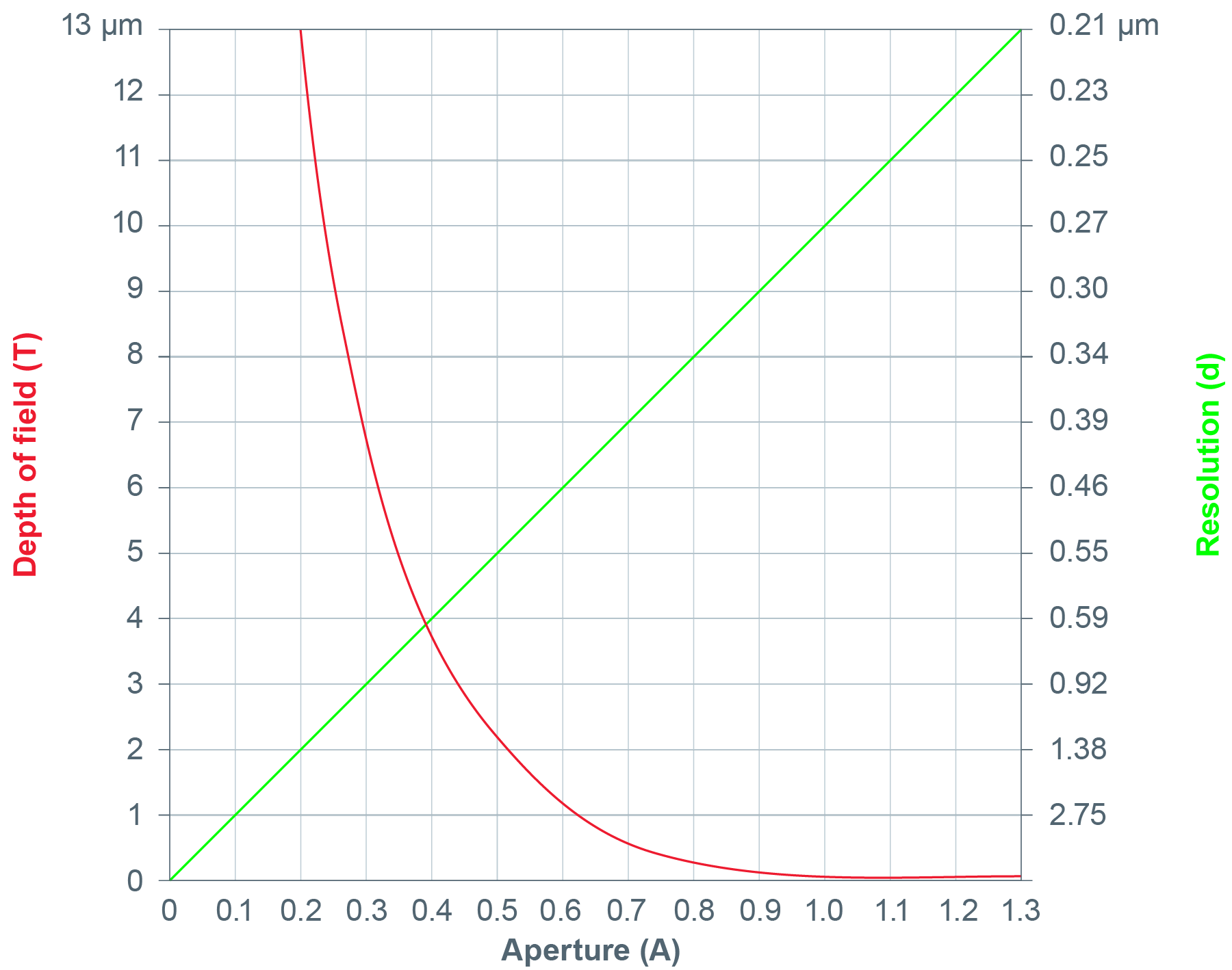
By using iris diaphragm objectives the numerical aperture of the objective can be changed. This is especially useful for widefield microscopy. If the iris diaphragm is closed, the numerical aperture and resolution are reduced but the depth of field is increased. If the iris diaphragm is opened again, the numerical aperture is increased, the resolution is increased, but the depth of field is decreased. An objective can also be used as a darkfield objective by narrowing the aperture.
Iris diaphragm objectives are labeled with the range within which the numerical aperture can be adjusted, for instance 1.4 – 0.7.
The physical relationship between aperture, resolving power and depth of field is shown in the graph. A small aperture produces low resolution but large depth of field. A high aperture means better resolution but less depth of field.
Linear correlation between aperture and resolution (green), respectively exponential correlation between aperture and depth of field (red)
Useful Sciencelab Stories
Collecting Light: The Importance of Numerical Aperture in Microscopy
Numerical aperture (abbreviated as ‘NA’) is an important consideration when trying to distinguish detail in a specimen viewed down the microscope. NA is a number without units and is related to the angles of light which are collected by a lens.
Beware of "Empty" Magnification
In the simplest case, an optical microscope consists of one lens close to the specimen (objective) and one lens close to the eye (eyepiece). The microscope magnification is the product of the factors of both microscope lenses. A 40x objective and a 10x eyepiece, for example, provide a 400x magnification.
Immersion Objectives: Using Oil, Glycerol, or Water to Overcome some of the Limits of Resolution
To examine specimens at high magnifications using the microscope, there are a number of factors which need to be taken into consideration. These include resolution, numerical aperture (NA), the working distance of objectives and the refractive index of the medium through which the image is collected by the front lens of an objective.
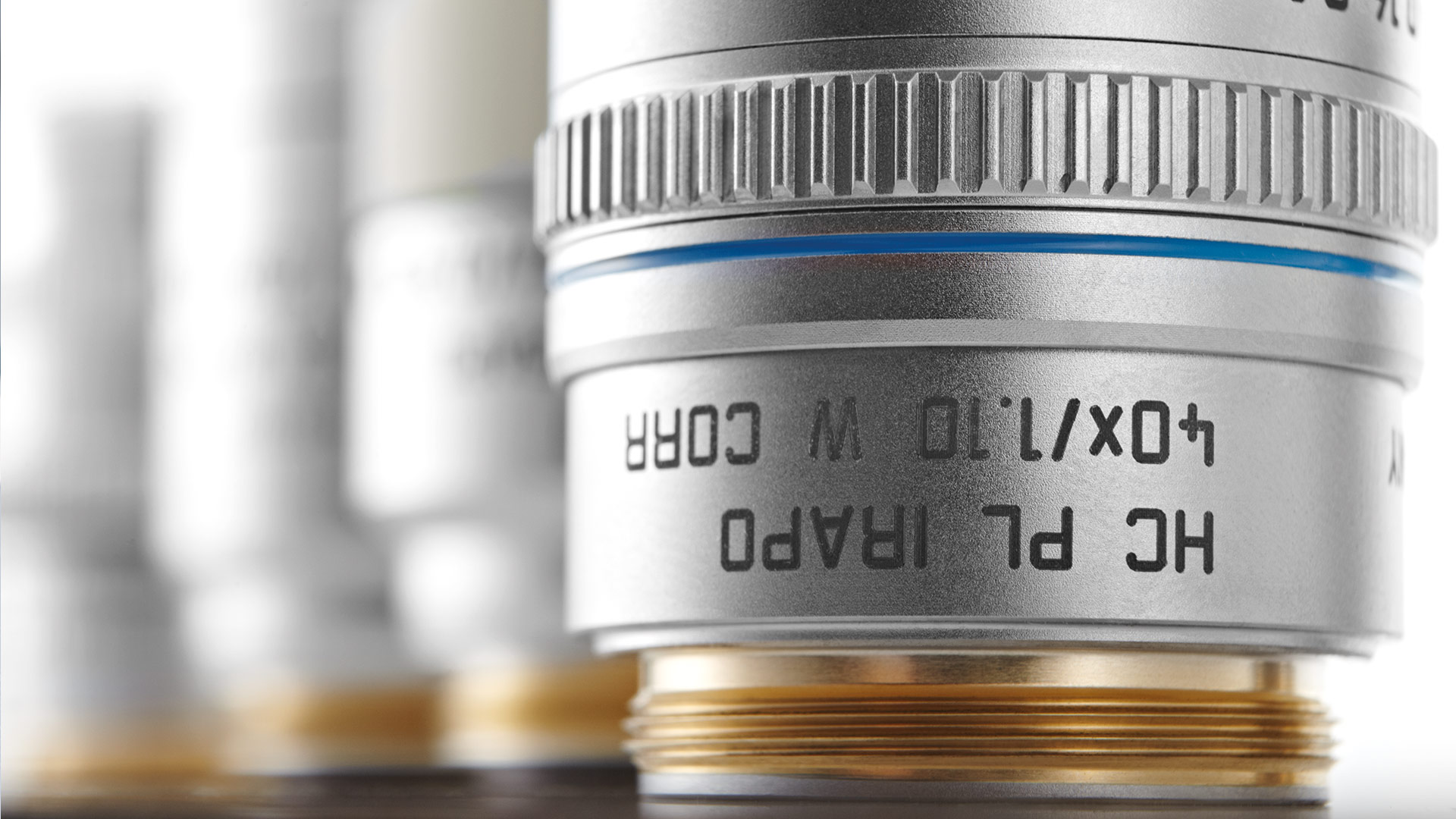
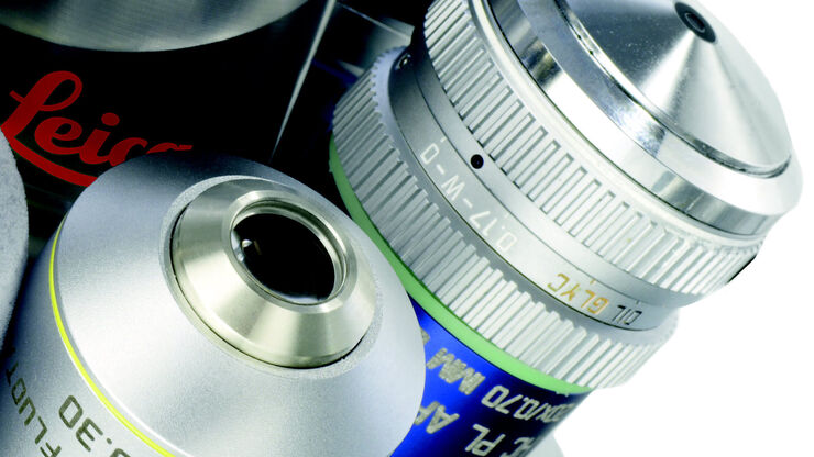
Correction Collar
The performance of high-resolution objectives is optimal when the refractive indices of the specimen and all intermediate optical media match the values for which the objective is designed. Changes in coverglass thickness and temperature as well as inhomogeneous, thick specimens introduce refractive index mismatches. This causes deterioration of the point-spread function, geometric distortion, and chromatic aberration. These effects limit penetration depth, contrast, and intensity of the microscope images.
Immersion oil traditionally has a refractive index close to standard crown glass. Oil immersion objectives are designed for the refractive index of this oil. They are optimal when working close to the coverglass or with samples embedded in a medium with a refractive index close to that of immersion oil. For samples with refractive indices deviating from this value special objectives are offered. Most common are water immersion objectives, and glycerol immersion objectives. Water and glycerol immersion objectives are very sensitive to changes in coverglass – introducing a changing thickness of a medium with refractive index mismatch – temperature, and deviations of the immersion medium or the sample itself. Therefore, water and glycerol immersion objectives with a higher NA have a correction collar to compensate for those differences.
The correction collar axially moves the central lens group and can be used to restore optimal image resolution and brightness. As manual adjustment of the correction collar requires time and experience and can disturb the sample, Leica offers water immersion objectives with a motorized correction collar.
CORR = Objective with correction collar
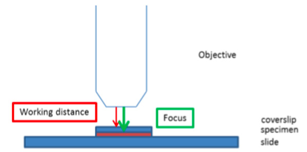
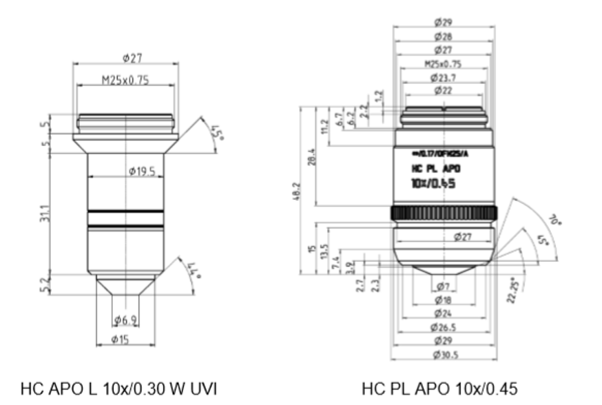
Working Distance
Free Working Distance
The free working distance (FWD) is an important application related feature of the objective. It is the distance between objective front end and coverslip surface facing the front lens (see Fig. ) The focus of the objective is exactly on the opposite side of the coverslip. I.e. The FWD is the distance you can focus into a sample, i.e. you can image inside the sample.
Extra-long Working Distance
The accessible sample area is often restricted by collisions of the objective with the sample holder, the edges of a multiwell plate or additional equipment, e.g. for electrophysiology or intravital imaging. Objectives with an extra-long free working distance allow to image also the edges of such samples without restrictions.
Deep tissue imaging by multiphoton excitation or in cleared tissues also requires large free working distance objectives to fully benefit from the optical advantages of those techniques. Here, the need for working distances of more than one milimeter are not uncommon. However, the numerical aperture of the objective still needs to be as large as possible to provide meaningful high-resolution images.
Leica Microsystems offers a range of objectives with exceptionally large free working distance for dry or water immersion. The water immersion objectives with extra-long working distances are also available with a large access angle and an inert ceramic front with minimal electric and thermal conductivity for electrophysiology.
L = Objective with extra-long free working distance
Water immersion objectives with extra-long free working distances
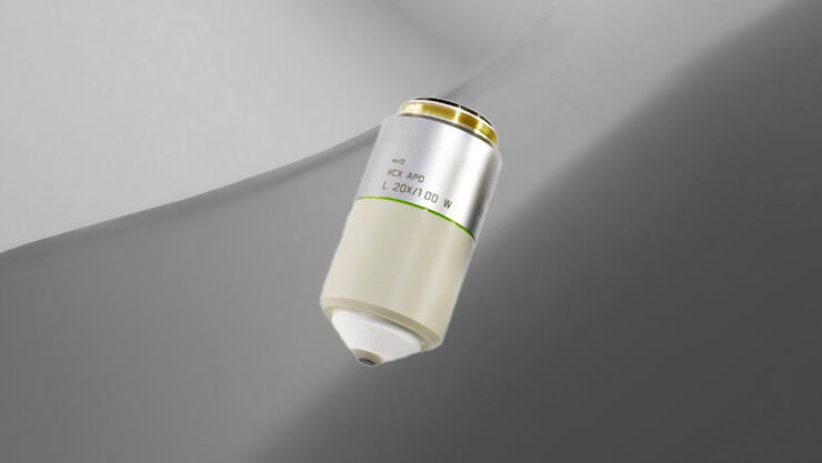
HCX APO L 20x/1.0 W with M32 thread for use on Leica DM6 FS and CFS, FWD: 2 mm
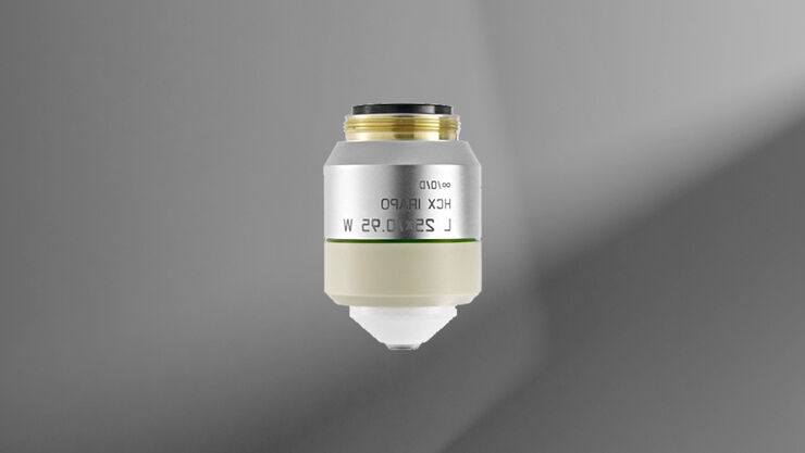
HCX IRAPO L 25x/0.95 W with M25 thread for use on all microscopes, FWD: 2.5 mm
HCX APO L U-V-I series, FWD: 2.2 – 3.6 mm
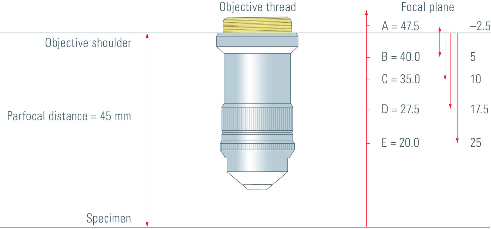
Back Focal Plane
Objectives by Leica Microsystems are defined by fixed back focal planes. The back focal plane code of the objective serves as a reference for selecting the matching objective-side DIC prism if needed. This has the advantage that several objectives can be used with one DIC prism.
A, B, C, D or E = Back focal plane of the objective
Matching Coverglasses
The coverglass is an important component of the optical path and should therefore meet the same standards of optical quality as the objective. High-quality objectives can only deliver their full potential if the corrected immersion medium and coverglass are used.
Dry, water and glycerol objectives are extremely sensitive to deviations in coverglass thickness. Objectives with a correction collar can be used to correct these deviations.
– | for use with and without coverglass |
|---|---|
0 | for use without a coverglass |
0.17 | for use with a 0.17 mm coverglass (DIN/ISO) |
1.8Q | for use with 1.8 mm quartz glass window on heating stages |
0-2 | for use with coverglasses of 0 - 2 mm thickness |
The available standard coverglass thicknesses are:
No. 1 | 0.13mm-0.17mm |
|---|---|
No. 1.5 | 0.16mm-0.19mm |
No. 1.5H | 0.17mm +/-0.005mm |
The coverglass type that is required for optimal results depends on the immersion medium and the numerical aperture (NA). Table 1 may be referred to as a general rule.
Coverglasses and immersion liquids:
| Immersion medium | With or without coverglass | Coverglass type 1.5 | Coverglass type 1.5 H |
|---|---|---|---|
| Air | NA < 0.30 | NA < 0.70 | NA > 0.70 |
| Water | NA < 0.60 | NA < 0.90 | NA > 0.90 |
| Immersion type G (glycerol) | NA < 0.80 | NA < 1.10 | NA > 1.10 |
| Immersion type N (Oil) | NA < 0.90 | NA < 1.30 | – |
| Immersion type F (Oil) | NA < 0.90 | NA < 1.30 | NA > 1.30 |
Table 1: Coverglasses and immersion liquids
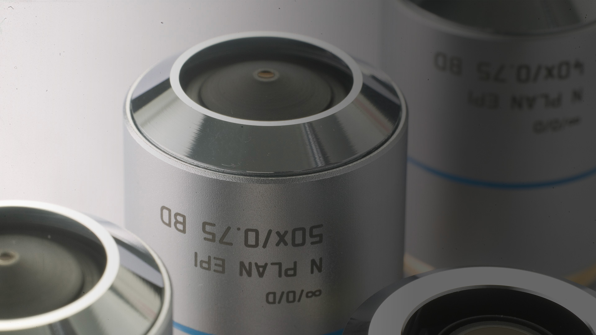
Contrasting Methods
Contrasting methods visualize phase shifts that are otherwise invisible to the human eye and thus enable observation of unstained living samples.
Objectives that are particularly suitable for specific contrasting methods are labeled accordingly.
BD | for brightfield/incident light darkfield |
|---|---|
PH | phase contrast objective |
RC | reflection contrast objective (only with DM R) |
P, POL | low strain, for quantitative polarization |
/ | not for incident light, except fluorescence |
LMC | modulation contrast objective (only with Leica DM IRB) |
Immersion Media for Objectives
For high resolving power, the numerical aperture (NA) of an objective needs to be greater than 1. This also requires an immersion medium with a refractive index greater than 1, i.e. other than air. Common immersion media are oil, water and glycerol. The immersion medium that has to be used with a certain objective is indicated on the objective.
Multi-immersion
Objectives suitable for the use with multiple immersion liquids, i.e. oil, water and glycerol, can be used with the optimal immersion medium for a large variety of samples.
Multi-immersion objectives:
- HC PL APO 10x/0.40 IMM CS
- HC PL APO 20x/0.75 IMM CS2
OIL | DIN/ISO standard immersion oil |
W | Water |
| GLYC | Glycerol |
IMM | Any other or more than one immersion media |
Refractive indices
All optically relevant elements (immersion medium, coverglass, sample) in front of the front lens of the objective have a major influence on the image quality. Ideally, the refractive index throughout all these optical layers should match the refractive indices the objective has been designed for. In reality, this is hardly possible as samples are often inhomogeneous, coverglass thickness is not that precise and the temperature changes during image acquisition.
These factors have to be kept in mind when choosing an objective and immersion medium for a certain application.
The higher the numerical aperture of the objective and the deeper the structures of interest inside the sample, the more important it is to match the refractive indices of the sample and the immersion medium. Different refractive indices lead to spherical aberrations and geometrical distortions of the structures. This results in a loss of contrast and definition, as well as structures appearing compressed or stretched.
Refractive indices of some important immersion media:
Cultured cells | 1.33 - 1.38 |
|---|---|
| Type F oil | 1.52 |
| Glycerol | 1.45 (21°C) - 1.46 (37°C) for Leica immersion liquid type G |
| Silicone oil | 1.41 |
| Water | 1.33 |
100% PBS pH 8,9 | 1.34 |
Mowiol | 1.46 |
| Canada Balm | 1.52 |
| CLARITY | 1.45 |
| BABB | 1.54 |
| Coverglass | 1.52 |
Oil
The refractive index of the Leica immersion oil type N and type F is 1.518 (at 23°C and 546 nm), i.e. the same as standard crown glass (n=1.518). For multicolor imaging, the dispersion of the immersion oil is also important. It is often referred to as the Abbe number and should match the Abbe number the objective was designed for otherwise, chromatic aberrations will occur. For example, Leica immersion oil type N has an Abbe number of 42.1 while immersion oil type F has an Abbe number of 46 which is optimal for fluorescence imaging. Type N is not suitable for fluorescence imaging.
Oil immersion objectives are ideal for samples that are in a medium that matches the refractive index of oil, i.e. classically fixed specimens embedded in resin, Canada balm or glycerol-gelatine, or are imaged close to the coverglass, i.e. less than a few µm. Further away from the coverglass the image brightness and resolution deteriorates quickly if the refractive indices are mismatched. For live cell imaging, i.e. imaging in an aqueous sample, we strongly recommend the use of water or glycerol immersion.
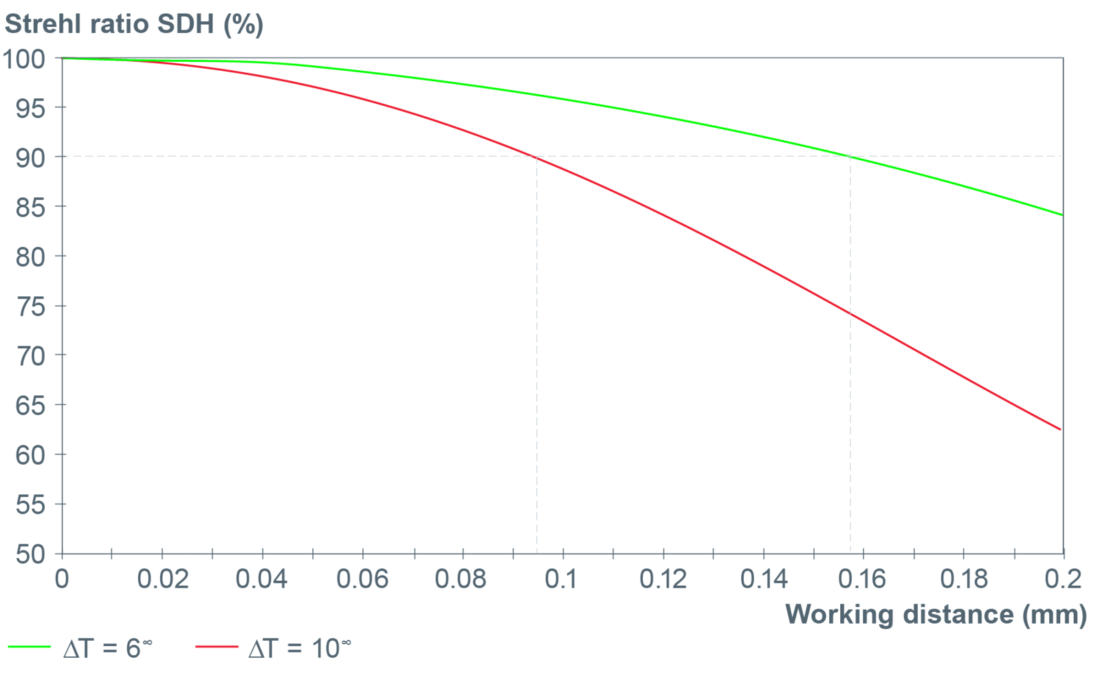
Oil immersion objectives with extremely high apertures only deliver their full optical performance in a relatively narrow temperature interval, as the refractive index of immersion oil greatly depends on the temperature. The larger the free working distance the thicker the oil layer, and therefore the larger is the impact of the temperature-related aberrations on the image quality. This temperature effect changes linearly with the free working distance but depends on the NA to the power of four.
For experiments at temperatures deviating from room temperature, water immersion is recommend as the refractive index of water is significantly less temperature dependent and can be compensated because many water immersion objectives have correction collars.
Water
Water immersion objectives are optimal for observing living cells in aqueous media as the refractive indices of the immersion medium and the sample are a closer match than for example immersion oil. However, water quickly evaporates at 37°C. The Leica Water Immersion Micro Dispenser automatically adds water during a running experiment to provide stable water immersion.
Glycerol
Today, most fixed samples like cultured cells, tissue slices of thick specimen as well as whole mount embryos are mounted in Mowiol, Vectashield or similar mixtures containing water and glycerol plus various chemicals, e.g. antifade and preservation substances. These media have refractive indices close to that of a 80%/20% glycerol/water mixture (n=1,45). Glycerol objectives are excellent for samples mounted in any medium with a refractive index close to 1.45 – 1.46.
Leica glycerol objectives offer a correction collar to adjust the optics to varying refractive indices coming from changes of the composition of the mounting medium, or from variations of coverglass thickness or temperature. Application Letter No. 17 (April 2004) gives a very detailed description of glycerol objectives and the effects of refractive index mismatches.
Glycerol Immersion
Objective for use with glycerol immersion:
- HC PL APO 63x/1.30 GLYC CORR CS2
- HC PL APO 40x/1.25 GLYC CORR CS2
- HC PL APO 40x/1.25 GLYC motCORR CS2
- HC PL APO 93x/1.30 GLYC motC STED W
Clearing Media
Voluminetric imaging of three-dimensional structures, whether in confocal microscopy, MP technology or lightsheet imaging, reaches the limits of what is possible after a few 100 µm. The reason for this lies in the different structures of a biological sample. For example, water has a refractive index (RI) of 1.33, proteins have an RI above 1.44 and lipids even above 1.45, which means that their interaction with the light passing through the tissue is different.
Light is scattered and absorbed by other structures, resulting in most tissue being non-transparent, which is particularly evident in larger tissue samples and deep imaging. To make these samples more transparent, different clearing technologies are applied. All tissue clearing techniques focus on "balancing" the refractive index to reduce inhomogeneities in light scattering without destroying the three-dimensional structure and without affecting the fluorochromes that may be present.
In order to achieve adequate clearing of hydrated samples still containing lipids. an RI of more than 1.45 must generally be achieved. At the same time, the objectives should have a sufficiently large working distance, a large field of view and, for spatial resolution, a high numerical aperture. Multi-channel multi-photon imaging also requires high performance in a spectral range from blue to near IR. A key to getting all these parameters right even at a depth of several millimeters is a control of the optical path length, i.e. a correction collar on the lens that allows adaptation to different refractive indices.
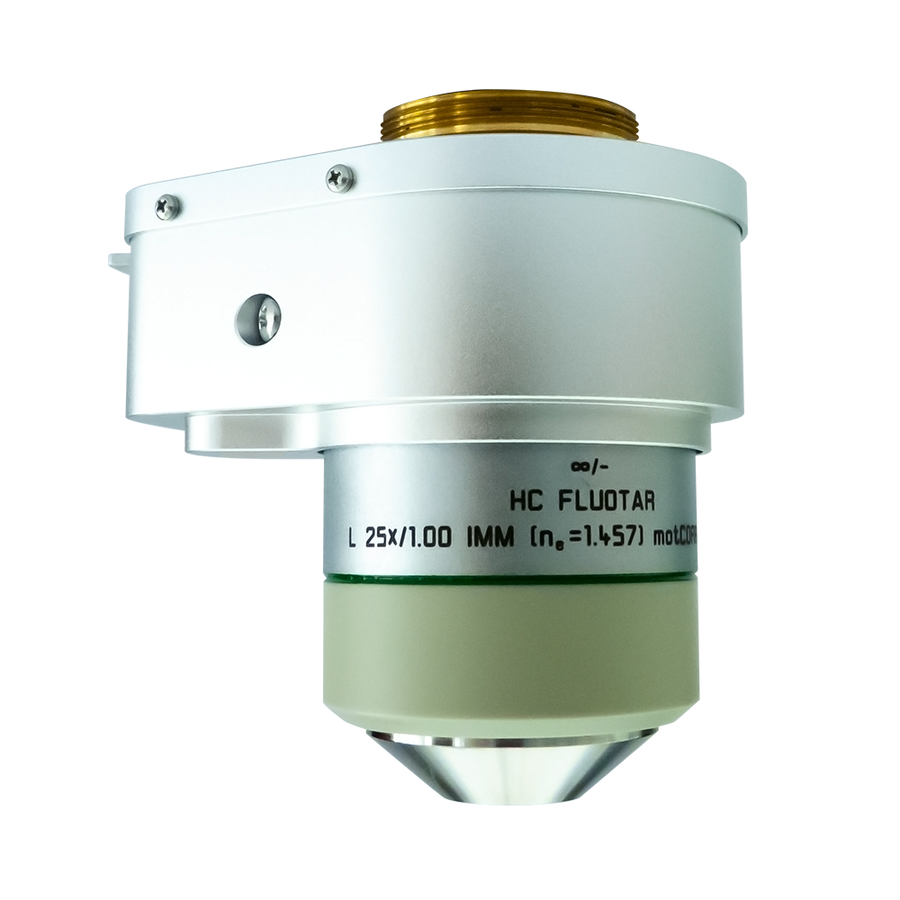
- HC FLUOTAR L 25x/1.00 IMM motCORR VISIR
In addition, some clearing methods use aggressive media such as BABB or ethyl cinnamate, which require specially designed objectives.
- HC PL FLUOTAR 5x/0.15 IMM DLS
- HC FLUOTAR L 16x/0.60 IMM CORR DLS
- HC FLUOTAR L 16x/0.60 IMM CORR VISIR
- HCX APO L20x/0.95 IMM
Objective Field Number
According to the standard ISO 19012-1, manufacturers of objectives now label their PLAN objectives with the correspondent objective field number “OFN”. This is a value for the flatness of the image over the field of view. The ISO standard defines field flatness using the Berek definition. The Berek formula (1927) delivers practical data for the visual depth of field (eyepieces).
Field of View and Object Field
A field of view of 25mm says that we have 25mm field in the intermediate image. To calculate the object field one divides 25mm by the objective magnification.
Example:
10x objective : 2.5mm object field
100x objective : 0.25mm object field
Diameter of Objectives
The diameter of an objective (with given parfocal distance) is a result of the specifications like numerical aperture, working distance and further technical design elements. Some objectives are specially designed for a slim shape, e.g. for a better access of patch capillaries to a brain section.
Related Articles

Factors to Consider When Selecting a Research Microscope

Infinity Optical Systems - From “Infinity Optics” to the Infinity Port

What is Empty Magnification and How can Users Avoid it
 where cyan indicates nuclei and magenta tight junctions.")
Rapid Check of Live Stem Cells in Cell-Culture Inserts set in Multi-Well Plates
Immersion Objectives
 of an image of two points where the distance between them corresponds to the Rayleigh criterion.")
Microscope Resolution: Concepts, Factors and Calculation

Eyepieces, Objectives and Optical Aberrations

Koehler Illumination: A Brief History and a Practical Set Up in Five Easy Steps

Collecting Light: The Importance of Numerical Aperture in Microscopy
Optimization of the Interplay of Optical Components for Aberration Free Microscopy
You have a need for sophisticated, highly specialized and unique lenses? You need small adjustments to existing lenses?
