EM TP
Sample Preparation for Electron Microscopy
Products
Home
Leica Microsystems
EM TP Automated Tissue Processor
Trust in the comparability of your results
Read our latest articles
EM Sample Prep Workflows & Uses
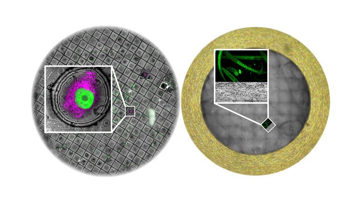
Researchers can consistently achieve high-quality, precise, and reproducible results when imaging samples with electron microscopy by using Leica sample preparation solutions. Our solutions support…
Fields of Application
Life Science Research
Leica Microsystems’ life science research microscopes support the imaging needs of the scientific community with advanced innovation and technical expertise for the visualization, measurement and…
EM Sample Prep Workflows & Uses
Researchers can consistently achieve high-quality, precise, and reproducible results when imaging samples with electron microscopy by using Leica sample preparation solutions. Our solutions support…
