EM TXP Cross-Sectioning Device
EM TXP is a mechanical cross-sectioning device that precisely downsizes samples for visual inspection. With EM TXP you can optimize sample pre-preparation to minimize ion beam milling time and ensure creation of mirror-like surfaces for light microscopy inspection.

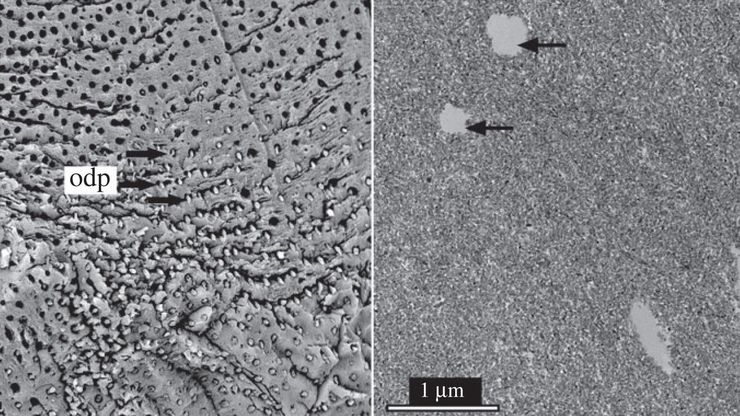
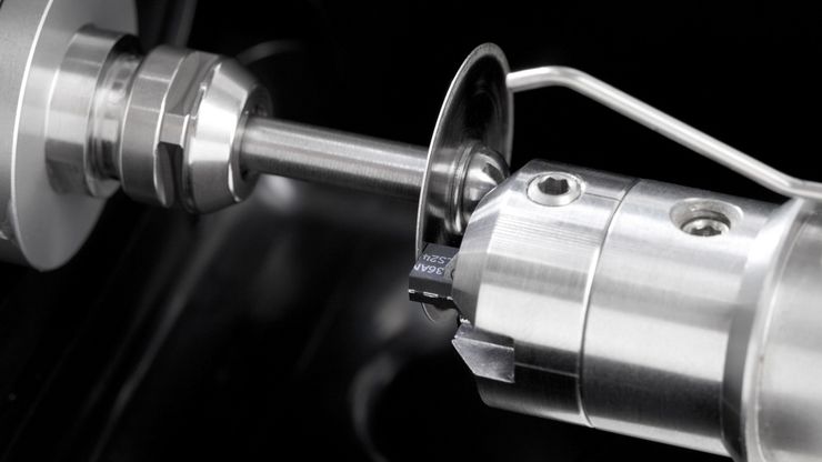
Buried structures prepared for broad ion beam milling
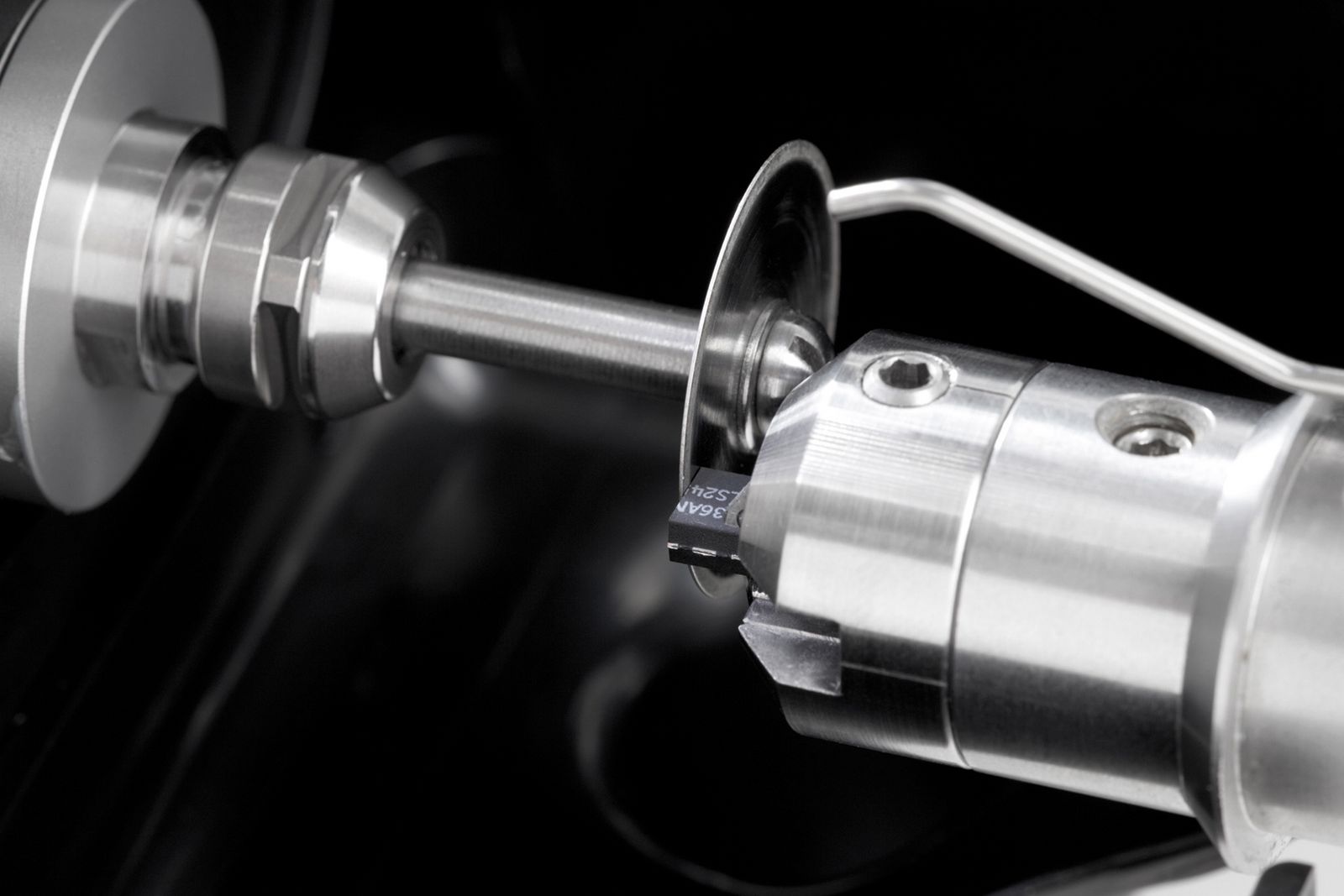
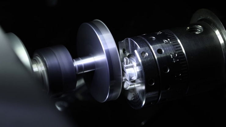
Multi-tool capabilities offer the right accessory for each process step, precisely honing in on your area of interest, and accurately positioning tool rotation and tilt angle.
Precise pre-cutting uncovers structures buried in the sample and shortens ion beam milling time by removing unwanted material.
Image adapted from Wang Yan et al., Comparative microstructural study on the teeth of Mesozoic birds and non-avian dinosaurs R. Soc. Open Sci.10230147 http://doi.org/10.1098/rsos.230147; https://creativecommons.org/licenses/by/4.0/
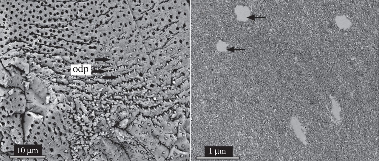
Fine-polished cross-section surface
- Prepare surfaces that meet your precision needs of light microscopy inspection.
- Precisely polish cross-section surface for visual inspection.
- Reveal your sample structure safely under stereo microscope observation.
Credits: Images Kurt Fuchs Photo Design. Photos taken by the Christiansen research group team in the Leica Microsystems Reference Laboratory at INAM/IKTS premises in Forchheim, Germany
Simply bring your sample through the workflow
Increase efficiency and reduce handling steps with sample support compatibility. Use the same holder for EM TXP and EM TIC 3X for the whole surface preparation workflow. Keeping consistent sample mounting and orientation through the entire preparation process decreases the risk of sample damage and misalignment.