Not all products or services are approved or offered in every market, and approved labelling and instructions may vary between countries. Please contact your local representative for further information.
With THUNDER Imagers you begin every experiment with confidence. They are your solution for advanced 3D cell culture assays, whether you want to study stem cells, spheroids, or organoids.
THUNDER Imagers feature innovative Leica technology Computational Clearing. It efficiently removes out-of-focus blur in real time, enabling the meaningful use of 3D specimens with camera-based fluorescence microscopes.
The high sensitivity of the system ensures low phototoxicity for physiologically relevant data sets. THUNDER systems offer a range of tools that assist you throughout your workflow from set up to data analysis.
THUNDER Imager Cell offers both speed and reliability for your 3D cell culture multiwell experiments. For example, when tracking development of organoids, the system's speed and reliability allows you to obtain excellent results.
Accurate multi-position time-lapse experiments are aided by:
Reliable focus drift correction with the Adaptive Focus Control (AFC)
Software autofocus that compensates for changes in specimen position
Reproducible Z-positioning with a precision of up to 20 nm (closed-loop focus)
The hardware triggered Quantum Stage moves rapidly and accurately to all positions with excellent repeatability (< ±0.25 µm).
Cultured VERO cells stained with STAR488 Vimentin (green), STAR580 Tom20 (yellow), and DAPI (blue). Sample courtesy Abberior GmbH, Göttingen (Germany).
Live cells contain a range of dynamic processes that require a system that can capture data with high temporal resolutions.
THUNDER Imagers only require a single fluorescence image to produce deblurred data. With THUNDER, you are only limited by the speed of your camera. THUNDER Imagers are offered with a range of cameras including the Teledyne Kinetix 22 allowing you to acquire data over 500 frames per second.
Mouse lung organoids derived from alveola stem and progenitor cells. Sample courtesy Dr. Pumaree Kanrai, MPI for Heart and Lung Research, Bad Nauheim (Germany).
Explore the features of the THUNDER Imager Cell with our interactive image map
Click on the + buttons to experience the technical highlights that support your fluorescence microscopy applications with high resolution imaging and optimal physiological conditions for your live cell cultures.
1
THUNDER Imager
Show details
Explore the features of the THUNDER Imager Cell with our interactive image map
Click on the + buttons to experience the technical highlights that support your fluorescence microscopy applications with high resolution imaging and optimal physiological conditions for your live cell cultures.
2
THUNDER Imager Cell (for basic screening)
Show details
3
THUNDER Imager Cell (for basic screening)
Show details
4
THUNDER Imager Cell (for live cell imaging)
Show details
5
THUNDER Imager Cell (for live cell imaging)
Show details
6
THUNDER Imager Cell (para escaneamento de placas de alto rendimento)
THUNDER Imager Live Cell was for me the perfect solution to get very high-quality images and also spend little time because it's just so fast to acquire each individual slide. By using THUNDER’s Large Volume Computational Clearing, I was able to remove this typical widefield haze and distinguish between background and signal. And that gave me the perfect starting point for my downstream analysis.
High throughput for better statistics and workflow efficiency
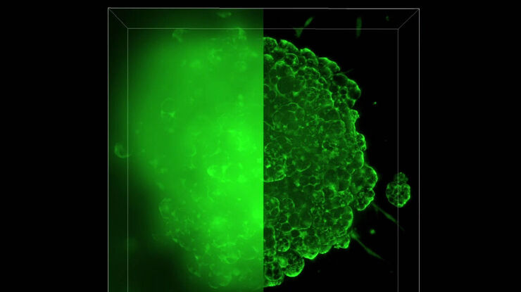
Automate your 3D cell culture assays to study next-generation disease models efficiently. THUNDER enables you to image large sample volumes, such as lung organoids, at high speed. In addition, the automation minimizes user interaction even for complex experiments.
THUNDER offers a range of solutions that allow you to acquire blur-free images of 3D samples with a single image.
This allows you to rapidly image over large areas, or at speeds of over 100 fps, at low light levels for physiologically relevant conditions, and gives you the freedom to set up a wide range of experiments.
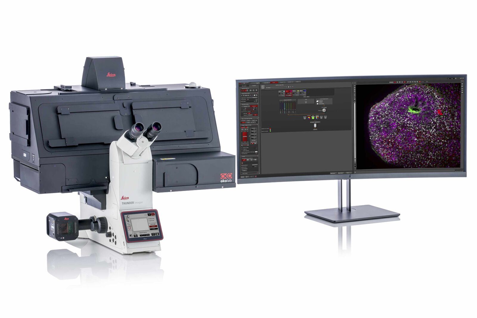
THUNDER Imager Cell is based on a fully motorized DMi8 microscope, Quantum Stage, highly sensitive K8 camera, and multi-line, high-intensity fluorescence LED light source. It is optimized for fast and precise multi-position, multi-channel imaging of 3D cell cultures.
Find the THUNDER Imager system that matches your needs
Whether you are looking for a dedicated high-end imaging system that excels in a given application, or a versatile solution for a lab running different kinds of assays with various samples, we’ve got you covered.
Here are some selected application examples that show you the strengths of THUNDER:
Mouse Retina
Show details
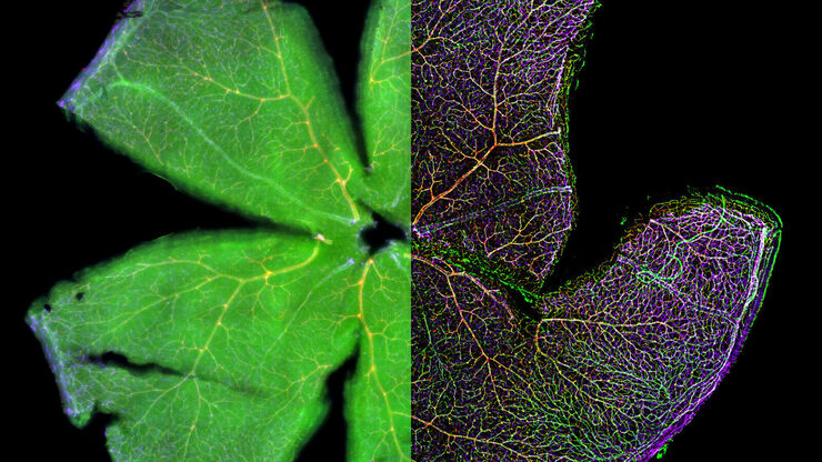
Reliable quantification of an entire mouse retina with THUNDER
Quantitative approaches to retina imaging analysis often require comprehensive descriptions of the retina morphology and function. Retina abnormalities as well as translational clinical applications need a reliable workflow to reproduce the transgenic target screening. Therefore, repetitive imaging of the morphology requires a system solution with accurate results which can be constantly reproduced. With the THUNDER Imager Cell you are able to clearly visualize the morphology and reliable count intracellular details, like single cell nuclei distribution in the retina.
With a THUNDER Imager Cell you have these advantages
Immediate blur removal helps you visualize more intracellular details
More utilizable depth with widefield approach
Reliable quantification
Ready-to-use for your particular workflow analysis
Control Swiss adult female mouse wholemount retina showing Iba1+ microglial cells (Alexa Fluor® 488 green-fluorochrome) and Brn3a+ retinal ganglion cells (Alexa Fluor® 594 red-fluorochrome). Image courtesy of Experimental Ophtalmology group, University of Murcia (Spain).
Brain Organoid
Show details
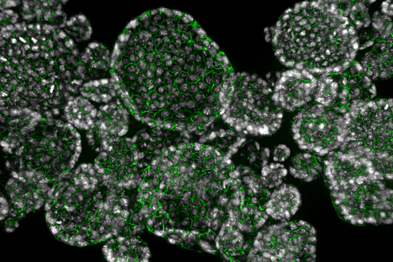
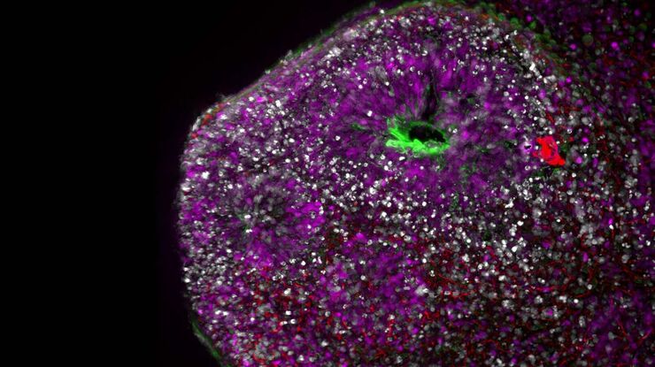
Visualization of brain organoids with a THUNDER Imager Cell
Brain organoids as novel model systems are used to investigate human brain development and disorders. These self-assembled 3-dimensional cytoarchitectures are often characterized via multiple transgenic marker imaging. Classical challenges in these workflows are quantifying dynamics of molecules in time besides keeping physiological conditions and reaching sample depth at low signal levels. Therefore, the THUNDER Imager Cell is suitable to study the development of organoids close to physiological conditions, as our LED light sources help to minimize photobleaching. Furthermore, the timed detection of even though low protein signal levels is possible without the need to change the carrier.
With a THUNDER Imager Cell you have these advantages
Possibility to observe your specimens in plastic bottom dishes makes your workflow more efficient
Capacity to detect low signals of molecules with high QE camera
Low bleaching and low sample perturbance with widefield approach and precise timed LED illumination
Nucleus (DAPI), p-vimentin (AF488), DCX (AF568), PAX6 (AF647). Uncleared human brain organoid (4 colors); Image courtesy by Atria Kavyanifar, M.Sc. (supervised by Prof. Dr. Lie, Prof. Dr. Winner) University Clinic Erlangen, Germany).
Explant Time-lapse
Show details
Low-phototoxicity in long-term live cell time-lapse imaging
Explant cell cultures are oftentimes difficult to conduct imaging experiments with, as they require a stable cell culture environment and low-phototoxic imaging conditions. This example of explant cell culture imaging from Dr. Laura Shankman at the University of Virginia shows how abdominal aorta cells were able to be stably imaged over a 48 hour period. The THUNDER Imager Cell provides a complete microscopy imaging system for minimally invasive and precise live cell imaging experimentation. Sensitive cell culture experiments can be efficiently conducted thanks to fast and High-Quantum-Efficiency camera options, accurate stages, tunable LED light sources, Computational Clearing to reduce out-of-focus blur in widefield images, and an easy to use LAS X software to automate imaging and analysis workflows.
Advantages of the THUNDER Imager Cell
Accurate live cell imaging experimentation allows tracking of fast cell movements
Low phototoxicity keep sensitive cell culture alive, even in long-term experiments
Speed-up your live-cell imaging workflow up to automated quantification and analysis
One week cultured explants of abdominal aorta imaged for 48 hours on gelatin coated #1.5 coverglass chamber slides. The mouse was genetically encoded with a smooth muscle cell specific tdtomato. After a transcription event, the smooth muscle cells excise the tdtomato and begin to express eGFP.

")