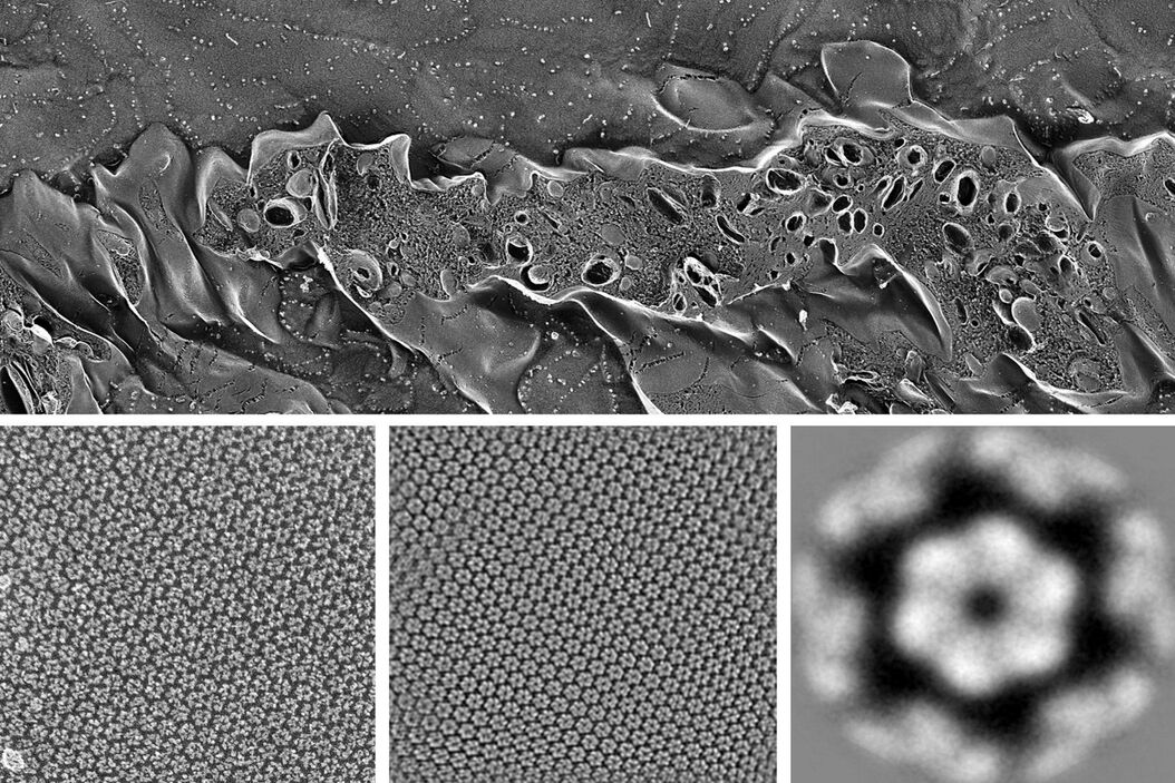
Freeze-fracture and freeze-etching are useful tools for studying flexible membrane-associated structures such as tight junctions or the enteric glycocalyx. Freeze-fracture and etching are two complementary methods for exposing membrane associated macromolecules, by means of sample vitrification for the preservation of targeted structure and then breaking a frozen specimen to reveal internal structures. Freeze etching is a follow-up step in which surface ice sublimates under vacuum to reveal further details. In these techniques, coating with metal or carbon enables the sample to be imaged in a cryo-SEM directly or in a TEM as a replica film. This pair of techniques is used to investigate cell organelles, membranes, layers and emulsions and is particularly useful for flexible structures that are not resolved by conventional or cryo-electron microscopy.
The ultimate resolution of these techniques is determined by how well the biological surface is replicated. Amorphous carbon replicas far better replicate biologic details than conventional metal coatings and using phase-contrast electron microscopy, provide unparalleled insight into these elusive cellular components.


