Falk Schlaudraff , Dr.

Falk Schlaudraff received his PhD from the University of Ulm and subsequently worked at Qiagen. He joined Leica Microsystems in 2011, initially as Product Manager for Laser Microdissection. Over the years, he took on responsibility for additional product lines and moved into leadership roles in product, application, and workflow management. Among other things, he oversaw innovations in Laser Microdissection (LMD) and played a key role in advancing Deep Visual Proteomics (DVP). Since January 2026, he has been leading the global Application Management Life Science Research team, focusing on advanced confocal, light sheet, and widefield microscopy.
 segmentation used in conjunction with LMD to increase discovery throughput.")
Biomarker Discovery with Laser Microdissection
Explore the potential of spatial proteomics workflows, such as Deep Visual Proteomics (DVP), to decipher pathology mechanisms and uncover druggable targets.
Altered protein expression, abundance, or…

Deep Visual Proteomics Provides Precise Spatial Proteomic Information
Despite the availability of imaging methods and mass spectroscopy for spatial proteomics, a key challenge that remains is correlating images with single-cell resolution to protein-abundance…
.")
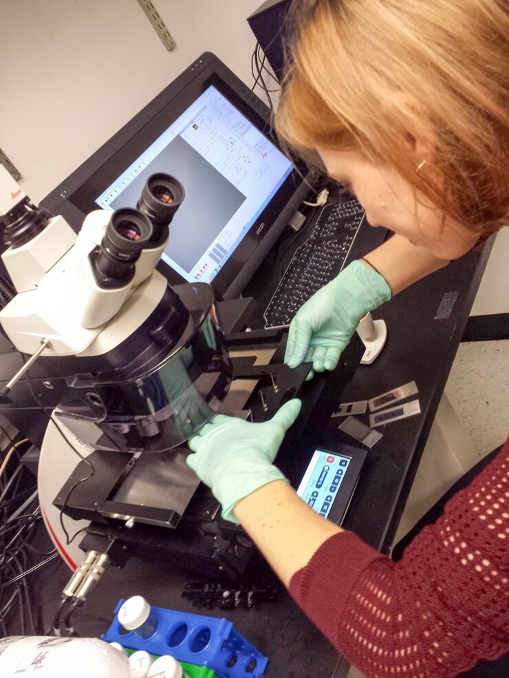
Neuron Isolation in Spatial Context with Laser Microdissection (LMD)
After Alzheimer’s disease, Parkinson’s is the second most common progressive neurodegenerative disease. Before the first symptoms manifest, up to 70% of dopamine-releasing neurons in the mid-brain…

RNA Quality after Different Tissue Sample Preparation
The influence of sample preparation and ultraviolet (UV) laser microdissection (UV LMD) on the quality of RNA from murine-brain tissue cryo-sections is described in this article. To obtain good…

The Cryo-CLEM Journey
This article describes the Cryo-CLEM technology and the benefits it can provide for scientists. Additionally, some scientific publications are highlighted.
Recent developments in cryo electron…

20 Years of Leica Laser Microdissection
Phenotype-genotype correlations are key for insight. From Eye to Insight is therefore fitting perfectly to Leica Microsystems and in particular to laser microdissection. Laser Microdissection, also…
Workflows & Protocols: How to Use a Leica Laser Microdissection System and Qiagen Kits for Successful RNA Analysis
Laser Microdissection (LMD) allows isolating individual cells or chromosomes and is a well established technique for sample preparation prior downstream analysis of the nucleic acid content via PCR or…
