
How does polarized light (POL) microscopy work?
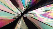
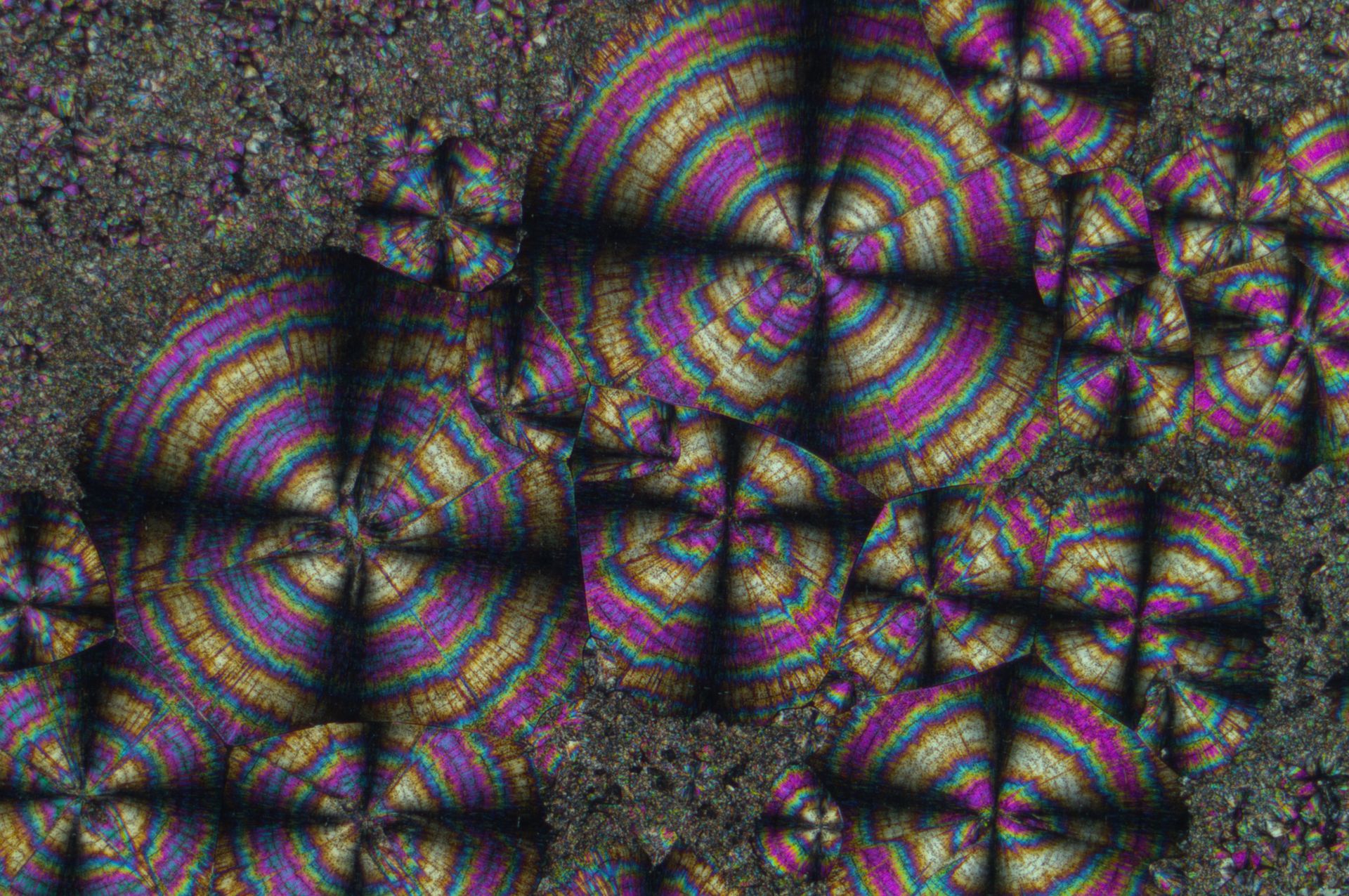
POL microscopy requires samples or specimens which are optically anisotropic, like birefringent materials. In these materials, the refractive index is not uniform and depends on the direction in which light passes through the material. When a sample has the property of birefringence, a ray of unpolarized light which passes through it can be divided into two or more rays due to differences in refraction. Examples of birefringent materials are calcite, boron nitride, cellulose, starch, glass with inclusions or under stress, or polymers under stress. Polarized light microscope images tend to show color effects which highlight birefringent structures.

Related Articles
The Polarization Microscopy Principle

Digital Microscopy in Earth Science

Polarized Light (POL) Microscope
Most people use brightfield illumination when imaging with a microscope. Unfortunately, it usually provides just a low-contrast image of many samples and specimens where few details can be distinguished. For life-science specimens, selective stains can be used to enhance the brightfield contrast, but they can be toxic to living cells. The refractive index, which depends on the speed of light in a medium, of a birefringent, optically anisotropic material depends on the direction in which light propagates through it. Polarized light can be used in microscopy to see color effects and the highlighting of structures in birefringent materials. Examples where polarized light microscopy is used are petrography, mineralogy, material structure characterization, asbestos analysis, coal analysis, quality control of glass, plastics, textiles, fibers, and electronic displays, and analysis of sub-cellular structures, like cellulose in the walls of plant cells and starch grains.
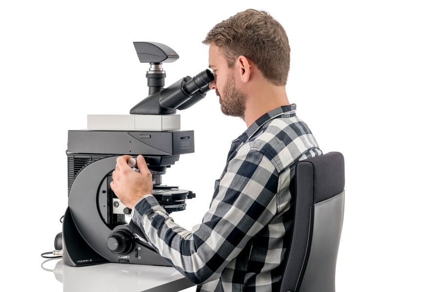
How do you set up a polarized light (POL) microscope?
In the setup of a POL microscope, the unpolarized light from the illumination source is polarized by a first polarizer. Afterwards, the now polarized light is focused on the sample by the condenser lens. If the specimen is birefringent or contains birefringent structures, the polarization plane of a portion of the light will be rotated by 90°. The image of the sample is magnified by the objective and hits the second polarizer. If this polarizer is twisted by 90° compared to the first polarizer, only light with a change in polarization after passing through the sample is able to reach the eyepieces or camera sensor and can be seen by observers. Sample structures which do not change the polarization of the light are not visible. For more information about polarization microcopy, refer to this article.
To avoid confusion, it is useful to point out that DIC (differential interference contrast) microscopes also use polarizers. However, in addition DIC requires a prism. More information about DIC microscopy is available in the related article below.

Applications where polarized light (POL) microscopy is used
POL microscopy is routinely used in the material and earth sciences to identify samples with birefringent structures, such as minerals, rocks, crystals, asbestos, and coal, based on their characteristic refractive properties and colors. For biology, POL microscopy is used for identification of cellulose in the walls of plant cells and starch grains. POL microscopy is also useful for quality control (QC) in various industries, such as glass and ceramics, plastics and polymers, textiles and fibers, and electronic displays.
 parallel polarizers and Right) crossed polarizers and a lambda plate.")
Related Articles
Ensuring Glass Quality with the Polarization Microscopy Advantage

Polarizing Microscope Image Gallery

FAQs Polarized Light (POL) Microscopy
A brightfield microscope uses ordinary brightfield illumination, which is non-polarized light, to observe the sample. A POL microscope uses polarized light to illuminate the sample.
Both POL and DIC microscopes use polarized light to observe the sample. However, DIC also requires a prism which disperses the polarized light into 2 distinct rays. If the rays experience different refraction or scattering from the sample, then different phase shifts occur. When these light rays reunite, they interfere and become elliptically polarized. This polarization can be changed into an amplitude shift which allows small height differences on the sample surface to be visualized.
The POL microscope was invented by the Scottish scientist Sir David Brewster in the early 1800s.
Yes, a POL microscope can be equipped with a camera for recording POL images observed with the contrast method. Make sure to find a camera that is good in low light settings.