The sections were imaged using a Tecnai Polara 300KeV (FEI, The Netherlands) microscope fitted with a 4K Gatan CCD camera. The magnification for the sections was 23K, with a defocus of -6um for the tomogram and -8um for the projection image, and the diffraction was done with a camera length of 930mm. The image in panel A is an average of the central 10 slices of a reconstruction done with the IMOD package (Kremer et. al., 1996), image processing software from a tomogram collected using the FEI software.
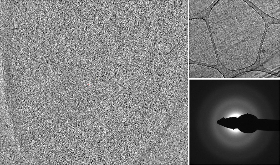
Left – optical slice from a tomographic reconstruction
Upper right – micrograph of a vitrified yeast cell
Lower right – diffraction pattern image
Related Articles
-

The “Waffle Method”: High-Pressure Freeze Complex Samples
This article describes the advantages of a special high pressure freezing method, the so-called…
Jul 08, 2025Read article -
.")
How Fluorescence Guides Sectioning of Resin-embedded EM Samples
Electron microscopes, including transmission electron microscopes (TEM) and scanning electron…
Jun 03, 2025Read article -

How to Save Time and Samples by Automated Ultramicrotomy
This article describes how 3D micro-CT data of a resin-embedded electron microscopy sample can be…
Apr 09, 2025Read article
Related Pages
-

Electron Microscope Sample Preparation
Excellent sample preparation is the prerequisite for first-class electron microscopy. Be prepared –…
Visit related page
