Key Learnings
- How to image multiple fluorescent labels with a single exposure using FluoSync – a new unmixing method
- How to use FluoSync to obtain 100% correlated labels without spatiotemporal mismatch, avoiding artefacts and false measurements
- Save time by eliminating the need to install filters in the lightpath
Speakers

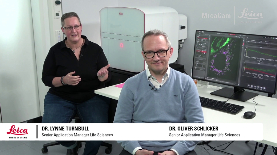
Dr. Oliver Schlicker, Senior Application Manager – Leica Microsystems
Oliver received his PhD in Neuro-Cell-Biology from the University of Heidelberg, Germany. After heading the Imaging Facility at the IZI in Stuttgart, Germany he joined Leica Microsystems Wetzlar in 2008 as Application Manager, responsible for Advanced Fluorescence Widefield Microscopy.

Dr. Lynne Turnbull, Senior Application Manager – Leica Microsystems
Lynne received her PhD in Sydney Australia in cardiac biophysics and undertook postdoctoral training in San Francisco and Melbourne. Upon moving to the University of Technology Sydney, Lynne established and managed the Microbial Imaging Facility. Lynne joined Leica Microsystems in 2021 as a Senior Application Manager and is based at the EMBL IC in Heidelberg.
The Experiment
Original broadcast date: 16th March 2022
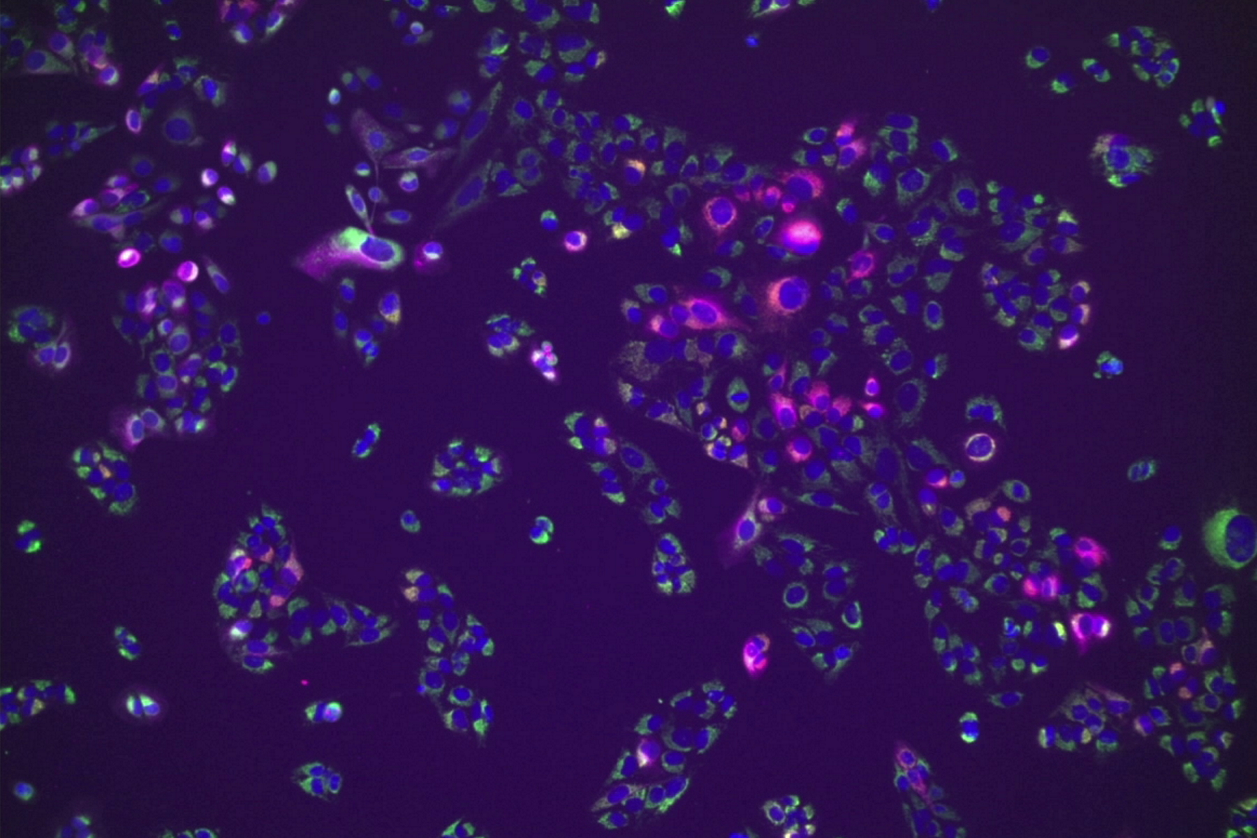
When studying multiple cell components over time, multiple fluorescent labels are typically used. Understanding the interaction of these components based on their spatiotemporal context is of interest to many researchers. Conventional imaging using one color filter after another cannot deliver this information with full precision as it uses sequential imaging of just one label at a time, rendering the outcome prone to crosstalk. The faster the biological events, the less precise the information you can acquire this way.
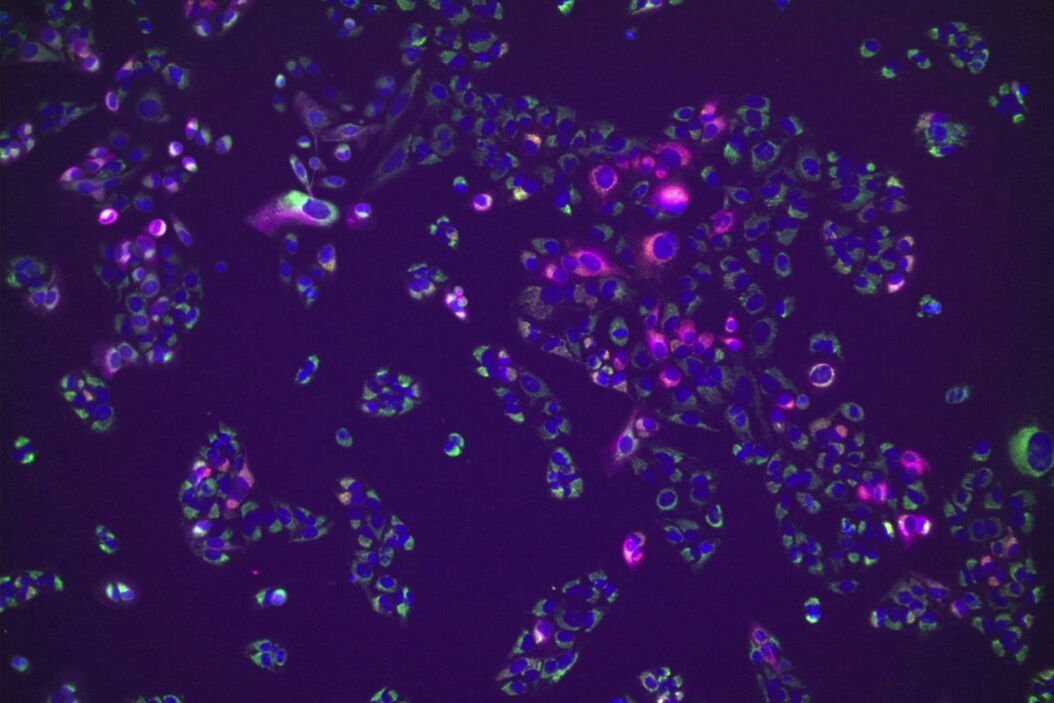
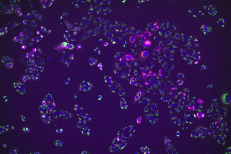
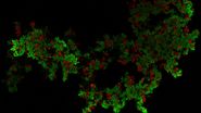
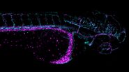
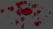
Mica, the world’s first Microhub, removes these constraints, and delivers absolute correlated labels without spatiotemporal mismatch, avoiding artefacts and false measurements. Thanks to the new unmixing method FluoSync, only a single exposure is required to image multiple fluorescent labels at the exact same point in time with full spatiotemporal resolution.
Setting up the experiment is also radically simplified when using Mica. Find out how much time you can save without having to install filters in the lightpath as you normally would with conventional imaging methods.
