
What to expect in the case study
Key Learnings
- Learn how automation, high-quality antibodies, and a proven workflow can deliver consistent results across samples
- See quantitative reproducibility data on a cohort of twelve slides
- Understand how the overall Cell DIVE solution can bring rock-solid results to your laboratory
About the case study
There is an ongoing crisis in reproducibility in scientific studies, and studies in spatial biology are no exception. Several factors can affect the reproducibility of experimental results. Antibody validation is an essential step in cancer research to ensure that the antibodies used are specific and reliable. These antibodies must maintain robust staining of samples, between lots, and over time. In recent years, automated systems are increasingly being used in cancer research to improve the reproducibility and efficiency of experiments.
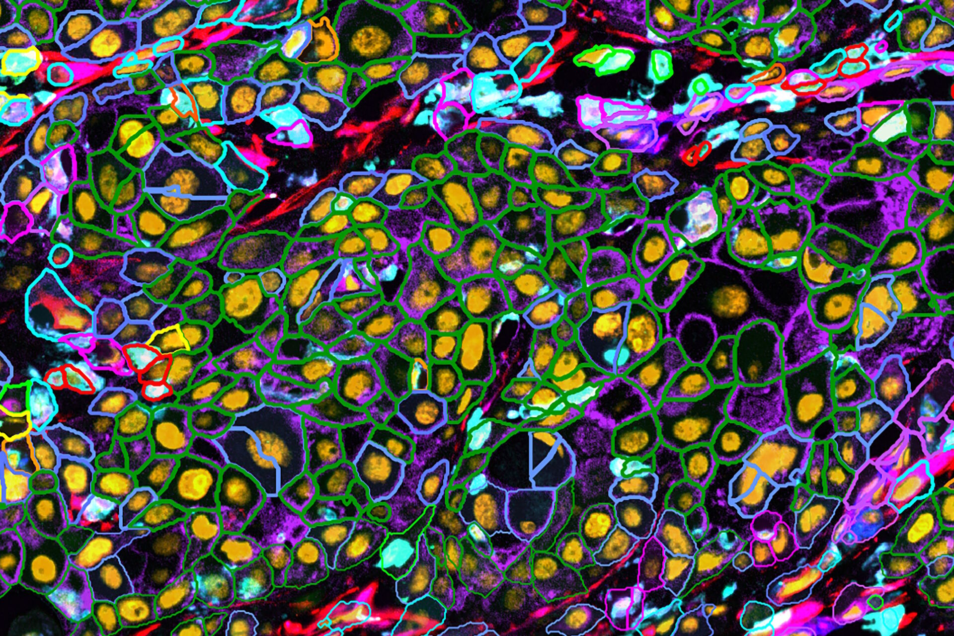
In this study, we test the reproducibility of the Cell DIVE multiplexed imaging solution. Cell DIVE with a BAB 200 automated system from Advanced Solutions Life Sciences (ASLS). We use validated antibodies from Cell Signaling Technology (CST). Cell DIVE allows probing and imaging of dozens of biomarkers on a whole tissue section. This is done with an iterative staining and dye inactivation workflow. At its core, Cell DIVE is designed to provide methods reproducibility, from tissue preparation, antigen retrieval, to sample imaging and slide storage. Cell DIVE is able to work with directly conjugated primary antibodies. This is yet another source of variability.
It's reassuring to know that conjugated antibodies from CST are rigorously validated to reduce variability. In addition, the use of recombinant antibodies, the consistent conjugate brightness, and antibody degree-of-labeling reduce lot-to-lot variability ensures reliable conjugated antibodies for spatial biology studies. The high-resolution imaging results are obtained by consistent round-to-round imaging, steady calibration, as well as reduction of human error with the BAB 200 liquid handling solution from ASLS. We also use adjacent tissue sections probed with distinct lots of CST antibody panels. Then, imaged in temporally separated batches using a Cell DIVE imager fitted with a BAB 200 liquid handler. Following robust analysis, we report the reproducibility findings across parameters. Methods and results reproducibility in spatial biology is essential for reliable insight generation, giving confidence in the quality of future studies aimed at improving patient outcomes.
")


