-b-poly(isoprene). Right: Poly(styrene)-b-poly(methyl methacrylate).")
Introduction
Polymers are foundational materials in modern science and industry, playing critical roles in applications ranging from packaging and textiles to biomedical devices and advanced electronics. Their versatility stems from the wide range of molecular architectures and morphologies they can adopt, which in turn govern their mechanical, thermal, and functional properties. Among these structural features, the lamellar organization—particularly in semicrystalline polymers—plays a pivotal role in determining performance characteristics such as strength, flexibility and barrier properties.
To fully understand and optimize these materials, it is essential to investigate their internal structure at the nanoscale. Transmission electron microscopy (TEM) is a powerful tool for this purpose, offering the resolution necessary to visualize the fine lamellar arrangements that are often inaccessible by other techniques. However, preparing polymer samples for TEM analysis presents unique challenges due to their soft, often heterogeneous nature. Ultramicrotomy, especially when performed under cryogenic conditions, enables the production of ultrathin sections that preserve the native morphology of the material.
In this study, we explore the use of the UC Enuity ultramicrotome from Leica Microsystems to section polymer samples under both room temperature and cryo conditions. We present high-resolution TEM images, proving essential role of the UC Enuity ultramicrotome in advanced sample preparation for polymer characterization.
Results
Sample preparation of polymers
As a first step for preparing polymers for ultramicrotomy one can divide the polymers into 2 classes: Rigid and soft. Rigid materials very often can be directly sectioned in the ultramicrotome, typically by using a diamond knife. For soft materials the most critical value is the glass transition temperature (Tg). It represents the temperature at which the polymer transitions from a hard, glassy state to a soft, rubbery state. In the context of ultramicrotomy sectioning, Tg is important because it helps determine the optimal temperature for sectioning, whether it can be performed at room temperature or under cryo conditions. Sectioning below the Tg can reduce deformation and compression, ensuring high-quality, ultrathin sections.
Nevertheless, many other factors influence the sectioning process: the processing, a potential blend or composite of materials, inclusions, nanoparticles, plasticizers and others. Furthermore, polymers can be stained as well (typically osmium tetroxide and/or ruthenium tetroxide), also influencing the brittleness, hence the behavior during sectioning.
Sectioning under room or cryo conditions
Typical polymer types, which can be sectioned under room temperature are e.g. polycarbonates, polymethylmethacrylates, stained polypropylene, stained high density polyethylene, epoxies, nylons and hard polyurethanes. Samples cut under cryo conditions are e.g. polypropylene (unstained), polyethylenes (unstained), rubbers, nylons, PVC, flexible polyurethanes, latexes, unstained ABS.
In the following we have chosen for room temperature poly(styrene)-b-poly(methyl methacrylate) and for cryo conditions poly(styrene)-b-poly(isoprene). The aim was to show the sectioning quality of the UC Enuity by visualizing the lamellar structure of both polymers in the TEM.
Room temperature sectioning of poly(styrene)-b-poly(methyl methacrylate)
Due to its hardness, the sectioning of poly(styrene)-b-poly(methyl methacrylate) was possible without cryo conditions. As the sample was synthesized in a small amount, the sample was embedded in an UV-curable resin for better handling. The resin adheres the sample to the sample holder within about 10 seconds under UV light.
The polymer was sectioned with a thickness of 60nm (Figure 1).
-b-poly(methyl methacrylate). Left: Sections on water in the knife boat during cutting guided by an eyelash. Right: Sections (white outlines) collected on grids with microgrid support.")
As the polymer was unstained the contrast was quite weak; nevertheless, the arrangement of the polymer fibers is already visible without phase contrast (Figure 2, left). Applying phase contrast enhanced the contrast drastically improving on the visibility of the regular polymer structure for further analysis. (Figure 2, right).
-b-poly(methyl methacrylate). Left: Standard TEM micrograph without phase contrast. Right: Same image position with phase contrast.")
Cryo sectioning of poly(styrene)-b-poly(isoprene)
In general, frozen or vitrified samples could be sectioned without resin-embedding. Nevertheless, as the synthesis was done on a laboratory level, the sample volume created here was not sufficient to hold it with standard sample holders. Hence, the sample was embedded in resin to increase the volume and to fix it onto the sample holder. The resin used was a UV curable resin, which hardens within seconds by UV exposure and the sample was mounted onto a cryo sample pin (Figure 3).
-b-poly(isoprene) under cryo conditions.")
Afterwards the sample was precooled in liquid nitrogen to speed up the cooling, as most polymer samples show poor thermal conductivity. However, depending on the polymer type other cooling methods may apply, as they can be disintegrated by rapid cooling in liquid nitrogen.
Then, the sample block was trimmed towards a block face and sectioned by the support of an eyelash, which can be subtly moved in parallel to the feed rate of the sectioning by the means of a micrometer screw. Sections of about 30 nm could be obtained and were then collected with a sucrose drop, transferred to an EM grid with holey carbon film and washed on a drop of water for TEM analysis.
Figure 4 shows an overview of the grid and the structure of the microgrid hosting the sections.
-b-poly(isoprene).")
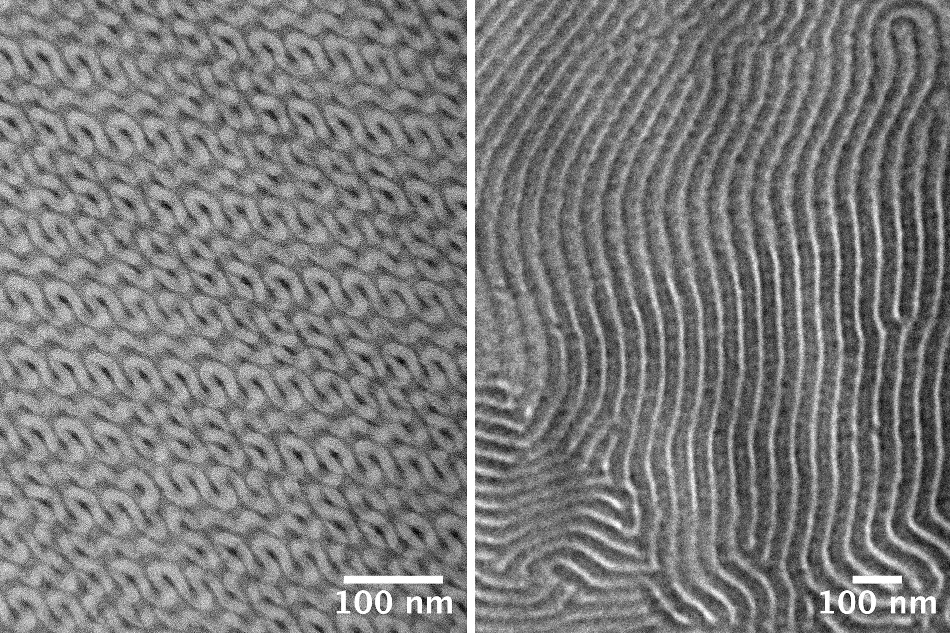
Due to the blistered structure of the supporting microgrid, several zones appear in the images depending on the position (without carbon support, with thin or thick carbon layer). Further magnification of the polymer on the unsupported grid region reveals insight into the structure (Figure 5).
-b-poly(isoprene). Left: 40.000x magnification. Right: 80.000x magnification. The coiled structure of the polymer becomes visible.")
The lamellar structure becomes clearly visible. This proves that the sections were well sectioned and collected. Furthermore, it shows that the fine morphology can be observed on a single section of 30-40 nm. Screenshots of the 3D reconstruction underline the orderly packed structure in 3D (Figure 6).
-b-poly(isoprene). Left: Oblique view. Center: Top view. Right: Top view rotated. The curled, regular structure is visible.")
Summary
This study evaluates the UC Enuity ultramicrotome from Leica Microsystems for preparing polymer samples under both room temperature and cryogenic conditions. The preparation process depends on the polymer's rigidity and glass transition temperature (Tg), which determines whether cryo sectioning is needed to avoid deformation.
Room temperature sectioning was demonstrated on poly(styrene)-b-poly(methyl methacrylate), which was embedded in UV-curable resin and sectioned at 60–90 nm. Despite being unstained, the polymer’s structure was visible, especially with phase contrast enhancement.
Cryo sectioning was applied to poly(styrene)-b-poly(isoprene). Due to limited sample volume, it was embedded in UV-curable resin and cooled with liquid nitrogen. Sections of 30–40 nm were successfully obtained and analyzed via TEM, revealing clear lamellar structures and 3D organization.
The results confirm that the UC Enuity ultramicrotome enables high-quality sample preparation for advanced polymer characterization.
Acknowledgements
We very kindly thank Prof. Hiroshi JINNAI for providing the samples. Special thanks to Akemi Kumagai for preparing the samples, sectioning and imaging (both Institute of Multidisciplinary Research for Advanced Materials, Tohoku, Japan).
, insulin SGs (orange), microtubules (red), nucleus (yellow), and plasma membrane (transparent).")


