Jan De Bock , Dr.

Jan De Bock

Jan studied biology and did his PhD in the field of olfaction, characterizing olfactory neurons in their response to odorants.

Jan has worked as a microscopy expert in different roles since 2003. He joined Leica Microsystems in 2011 as a product specialist for confocal microscopy. In 2017, he became member of the newly formed Workflow and Application Team responsible for correlative workflows, in particular involving sample preparation and imaging under cryogenic conditions.

Projection of a confocal z-stack. Sum159 cells, human breast cancer cells kindly provided by Ievgeniia Zagoriy, Mahamid Group, EMBL Heidelberg, Germany. Blue–Hoechst - indicates nuclei, Green–MitoTracker mitochondria, and red–Bodipy - lipid droplets

New Imaging Tools for Cryo-Light Microscopy

New cryo-light microscopy techniques like LIGHTNING and TauSense fluorescence lifetime-based tools reveal structures for cryo-electron microscopy.
Correlation of markers in the LM and the FIB image.

How to Target Fluorescent Structures in 3D for Cryo-FIB Milling

This article describes the major steps of the cryo-electron tomography workflow including super-resolution cryo-confocal microscopy. We describe how subcellular structures can be precisely located in…
HeLa Kyoto cells (HKF1, H2B-mCherry, alpha Tubulin, mEGFP). Left image: Maximum projection of a z-stack prior to ICC and LVCC. Right image: Maximum projection of a mosaic z-stack after ICC and LVCC.

How to Improve Live Cell Imaging with Coral Life

For live-cell CLEM applications, light microscopy imaging is a critical step for identifying the right cell in the right state at the right time. In this article, Leica experts share their insights on…
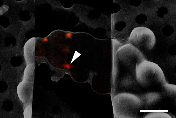
Cryo FIB lamella - Overlay of SEM and confocal fluorescence image. Target structure in yeast cells (nuclear pore proteine Nup159-Atg8-split Venus, red) marked by an arrow. Scale bar: 5 µm. Alegretti et al.,  Nature 586, 796-800 (2020).

Targeting Active Recycling Nuclear Pore Complexes using Cryo Confocal Microscopy

In this article, how cryo light microscopy and, in particular cryo confocal microscopy, is used to improve the reliability of cryo EM workflows is described. The quality of the EM grids and samples is…

Crystal Clear Cryo Light-microscopy Images

This article describes how computational clearing of cryo light microscopy images improves the identification of cellular targets for cryo electron-microscopy.
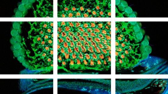
Scheme of a 2D mosaic scan. Drosophila melanogaster (eye section)

Mosaic Images

Confocal laser scanning microscopes are widely used to create highly resolved 3D images of cells, subcellular structures and even single molecules. Still, an increasing number of scientists are…
Scroll to top