
Enhancing vascular blood flow visualization: The need for fluorescence solutions
When operating under white light, neurosurgeons can clearly see tissues and vessels, but they cannot visualize blood flow. Full visualization of blood flow and anatomy during cerebrovascular procedures is critical for confident assessment and decisions. In the past, you could only view flow by pausing surgery and watching the black and white near-infrared (NIR) fluorescence video, which meant losing depth perception and anatomical detail. This forced surgeons to switch between the microscope’s white light view and the NIR view sequentially, having to observe and mentally reconcile information from both views to gain a comprehensive understanding. This not only took time and effort but also increased the risk of overlooking critical details due to the difficulty of having to recall all spatial features.
Furthermore, NIR fluorescence intensity is affected by working distance and magnification. Surgeons can therefore only compare intensity within the same field of view at a particular step of surgery, not across different steps, thus making real-time, comparative analysis challenging. Leica Microsystems has overcome these challenges with the GLOW800 3D Augmented Reality (AR) technology. GLOW800 directly addresses the limitations by fusing the natural color white light microscope view with a digitally enhanced visualization of the blood flow, highlighted in a pseudo-color, all in a single augmented real-time view. This Augmented Reality solution allows surgeons to simultaneously observe the intricate details of vessels and tissues and the blood flow, both with 3D depth perception, live on a large 3D 4K monitor or with the Leica 3D headset MyVeo. This eliminates the need for mental reconstruction, providing a seamless and comprehensive understanding of the operating field, aiding surgical precision and efficiency during critical neurovascular procedures.
Advantages of using the GLOW800 AR fluorescence application for surgery
Visualization with GLOW800 AR fluorescence supports each step of a surgery, for example during aneurysm clipping, it helps:
- Assess clip placement and aneurysm occlusion
- Check if all branches proximal and distal to the clipped aneurysm are perfused and whether there is orthograde filling of the blood vessels
- Confirm the clip has not caused any compromise of surrounding blood vessels, such as kinking or partial obstruction
- View blood flow without interrupting the surgical workflow
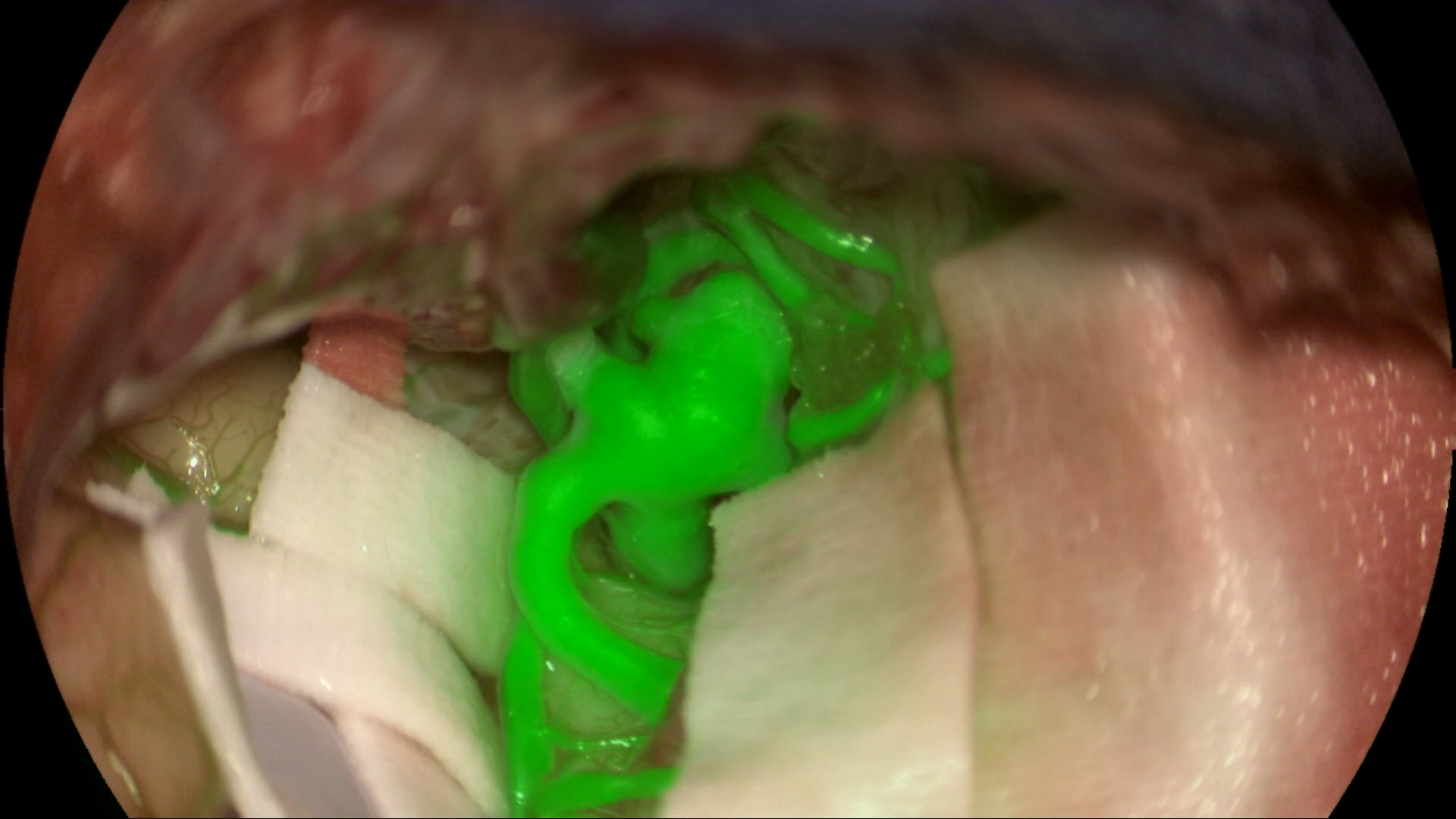
Aneurysm clipping under white light with GLOW800. The blood flow is highlighted, and it allows surgeons to better assess the aneurysm clip and blood flow post clipping. Video courtesy of Dr. Jacques Guyotat, Hospices Civiles de Lyon, France.
How does the GLOW800 AR fluorescence application work in detail?
The GLOW800 AR fluorescence application involves combining information from different imaging processes and wavelengths. The white light reflectance imaging illumination consists of visible light with a wavelength of approximately 400-700 nm, and the ICG fluorescence excitation illumination consists of near-infrared light, approximately 700-790 nm.
All light above 790 nm is filtered out to avoid interference with the fluorescence emission signal. The GLOW800 multispectral sensor employs filters to allow independent measurements in four spectral bands: red, green, and blue for reconstructing the white light reflectance image, and NIR emission at 835-880 nm for visualizing ICG fluorescence.
The spectral graph (figure 1) below illustrates the light wavelengths which are and are not visible to the human eye. By exciting the fluorophores in the non-visible range, they become ‘visible’ to the human eye through the GLOW800 filter.
.")
| Fluorescence excitation | 790 nm |
| Fluorescence signal | 835 nm |
Table 1
The GLOW800 application uses a video processing unit (VPU) to acquire, process and display the data of two video streams from cameras embedded in the microscope optical head.
Customizing NIR fluorescence imaging with pseudo coloring
The standard image of NIR fluorescence is typically black and white. However, surgeons can choose a pseudo (false) color (as shown in figure 2 and table 2 below) based on personal preference for observing the emission fluorescence signal. The color image from white light illumination is constructed using a 'debayering' (or demosaicing) process, which interpolates color values from the filter array output of the GLOW800 sensor chip.

| Color Option | Azure | Blue | Dark Cyan | Green | Magenta |
| RGB Value | 255-000-255 | 127-000-255 | 000-127-255 | 000-221-221 | 000-255-000 |
Enhancing luminance uniformity with homogenization filters
To overcome spatial inhomogeneities in illumination and imaging optics, a digital homogenization filter is applied to each incoming frame. This filter normalizes the measured signal by applying a pixel-wise linear correction based on microscope settings (working distance, magnification, iris, illumination), as illustrated in Figure 3 below. Digital homogenization compensates effectively for center-weighted brightness variations. The degree of homogenization can be manually adjusted by the user, ranging from 0% to 100%.
Compensating for brightness variations intensity scaling
The brightness of acquired images decreases with increasing working distance and magnification, as well as with decreasing light intensity. To compensate for this decrease, a scale factor is applied to the fluorescence image. The intensity adjusts the fluorescence contrast, brightness, and transparency in relation to the object details. The intensity values range from 0% (fluorescence just visible/object is dominant) to 100% (fluorescence intensively visible and dominant).
The power and potential of multispectral imaging applications by Leica Microsystems
The GLOW800 and GLOW400 clinical applications of the GLOW AR platform are based on digital spectral detection augmenting structures and tissues with multiple spectral bands. The sophisticated imaging sensors and algorithms of GLOW AR acquire, optimize, and combine multiple spectral bands of light. The result is natural or bright coloring of the anatomy and accurate representation of fluorescence intensity in a high-definition 3D image.
Whether you need to see vascular flow augmented in your white light microscope view or see clearer anatomical details surrounding the fluorescent-marked tumor, the clinical applications of the GLOW AR platform provide you and your entire team with real-time 3D AR views for confident, precise and well-informed surgical decision-making.
The GLOW AR platform is currently available on the ARveo 8x microscope.
 case.")