
Robustness of microscopes to environmental conditions
The performance of optical instruments can be affected during their use by a number of environmental factors, for example, high temperature and humidity typically found in tropical climates. These factors depend on the place of use, e.g. laboratory, workshop, factory, etc., and the geographic location. Individual factors can cause a variety of effects on instrument performance. In general, the manufacturer attempts to ensure that the instrument is robust enough to withstand a large range of environmental factors and that its performance will be maintained successfully during its lifetime.
The performance robustness of an instrument can be assessed by exposing it to a range of simulated environmental factors under controlled laboratory conditions. Accelerated testing is often exploited where the severity of conditions is increased at regular intervals to obtain meaningful results in a relatively short period of time.
The basis of the international standard ISO (International Organization for Standardization) 9022, which concerns environmental test methods for optics and optical instruments, is the assessment and comparison of the performance of optical instruments exposed to varying environmental conditions. ISO 9022 describes in detail a number of laboratory tests which reliably simulate a variety of different environments, where the tests are based largely on IEC (International Electrotechnical Commission) standards [1,2]. The effects of mold and fungus are tested in ISO 9022-11; procedures for optics and optical instruments, environmental test methods, part 11: mold growth [3]. ISO 9022-11 specifies methods for testing the robustness of optical instruments to the effects of mold and fungus growth, mainly a concern in hot, humid environments, such as tropical or sub-tropical regions of the world, but can also be a problem during the summer seasons globally.
Normally, these tests are performed with representative samples, e.g. mounted optics, material specimens, or surface coatings on such representative samples. The purpose of the testing is to investigate how the optical, chemical, mechanical, and electrical performance characteristics of the samples are affected by mold and fungus growth and their metabolic waste products, i.e. enzymes and acids.
ISO-9022 test on Leica microscopes
To verify that the objective lenses, as well as other components, of Leica microscopes are resistant to the growth of mold or fungus, rigorous tests in accordance with ISO standards have been performed by Division 4.1 ("Biodeterioration and Reference Organisms") of the DAkk (Deutsche Akkreditierungsstelle) accredited BAM Federal Institute for Materials Research and Testing in Berlin [4].
The test methods followed the ISO 9022-11 procedure (optics and optical instruments, environmental test methods, part 11: mold growth) [3] mentioned already above. Microscope parts tested include objective and condenser lenses, eyepiece lenses and tubes, a digital camera, among others. The testing involved 10 species of mold/fungus under the following conditions: a temperature of 29 °C (84 °F) and a relative humidity (RH) of 96%. The entire test occurs over a 4-week period.
Spore concentrations (over surfaces) inside the testing chamber were in the range of 12,000 to 18,000 spores/cm2 (120 to 180 million spores/m2). In comparison to what are considered as acceptable spore concentrations for indoor environments [5, 6], the spore concentrations in the test chamber are approximately 700,000x to 1,000,000x higher. For a typical range of values measured for outdoor environments [5,7,8], the spore concentrations in the test chamber are 1,000x to 10,000,000x greater. Figure 1 is a video showing the misting process used to spread the mold and fungus spores all over the inside of the chamber at the start of the test.
Control experiments using filter paper and cotton samples were done simultaneously under the same conditions as for the microscope parts. After 1 week, both the paper and cotton control samples were completely covered from the growth of mold and fungus, demonstrating the severity of the test conditions.
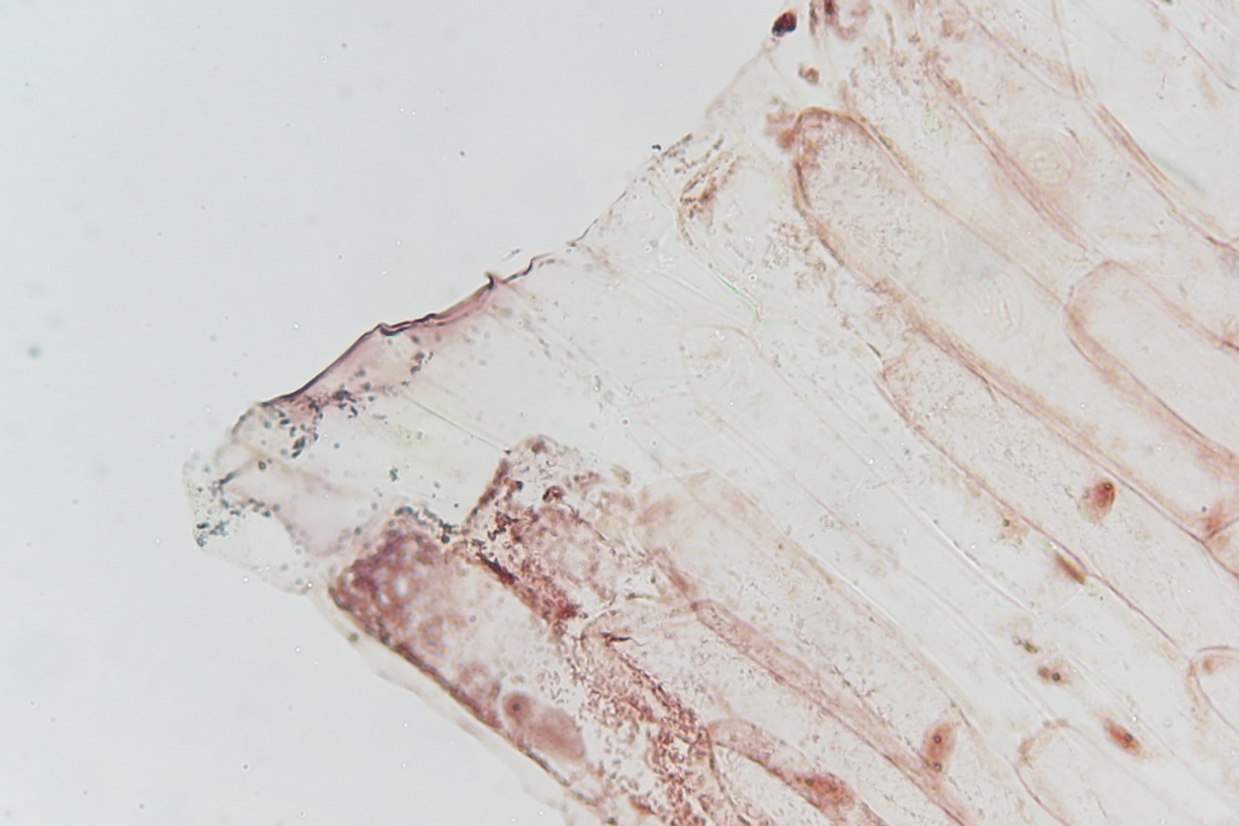
Final results for some microscopes of the Leica educational series show that all parts received a grade rating of 0, 1, or 2. A grade of 2 or less indicates successful resistance to mold and fungus growth . More specific details can be found in the clarification letter issued after receipt of the final test results from BAM [9]. Examples of an image acquired of an onion flake with a basic Leica compound microscope (40x achromatic objective and 10x eyepiece) both before and after the ISO 9022-11 test are shown below in figure 2. The images were recorded by a digital camera viewing through an eyepiece.

Summary and conclusions from the test results
The Division 4.1 of the BAM Federal Institute [4] performed a rigorous mold and fungus resistance test, in accordance with the ISO 9022-11 procedure (environmental test methods for optics and optical instruments: mold growth), for various Leica microscope parts, including objective lenses.
The final results of the rigorous mold and fungus test clearly confirm that the main components of the Leica educational series of microscopes successfully resist mold and fungus growth [9]. Therefore, mold and fungus should have no effect on their performance .
In addition to the ISO testing, silver (Ag) based coating additives, such as AgTreatTM, are used with main touch-point plastic and painted components of some Leica microscopes in order to inhibit the growth of bacteria .
Acknowledgements
We would like to thank Dr. Ina Stephan of the Biodeterioration and Reference Organisms Division of BAM Federal Institute for Materials Research and Testing in Berlin [4] for supplying the video of misting with a solution of mold and fungus spores inside the test chamber (refer to video 1).