Access to Webinar
Access the webinar@BiteSizeBio: THUNDER Imagers Imaging Workflows
Related Articles
-
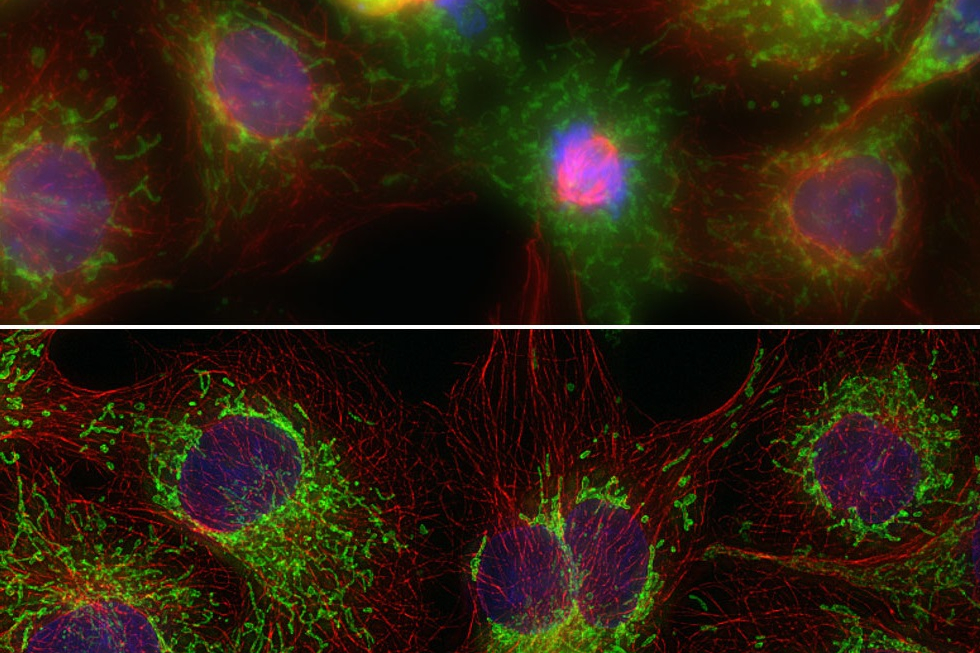
, tubulin with Cy5 (red), and nuclei with DAPI (blue). Image courtesy of Dr. Günter Giese, Max Planck Institute for Medical Research, Heidelberg, Germany.")
Overview of Fluorescent Dyes in terms of Applications and Properties
An introduction to commonly used fluorescent dyes and an overview of their characteristics are given…
Mar 16, 2026Read article -

Factors to Consider When Selecting a Research Microscope
An optical microscope is often one of the central devices in a life-science research lab. It can be…
Dec 16, 2025Read article -

Technical Terms for Digital Microscope Cameras and Image Analysis
Learn more about the basic principles behind digital microscope camera technologies, how digital…
Dec 14, 2023Read article
Related Pages
-

THUNDER Imaging Systems
To answer important scientific questions, THUNDER Imaging Systems enable you to obtain a clear view…
Visit related page
