Murine esophageal organoids (DAPI, Integrin26-AF 488, SOX2-AF568) imaged with THUNDER Imager Cell. Courtesy of Dr. F.T. Arroso Martins, Tamere University, Finland.

Advanced Imaging Solutions for Biopharma

Advanced imaging solutions empower biopharma researchers to uncover critical insights in drug discovery, quality control, and understanding complex biological systems. Leica microscopy solutions combine innovation, user-centric design, and reliable data integrity to support breakthroughs and consistent research outcomes.

Simply get in touch!

Our imaging experts are here to help with advice on solutions for the biopharmaceutical industry.

Co-culture of CAR-T cells and breast cancer organoids. CAR-T cells labeled by CD3 (green). Breast cancer organoids labeled by membrane marker B7H3 (blue). DNA labeled by SiR-DNA (red). Image acquired using STELLARIS. Image courtesy of Dr Ravian van Ineveld and Prof. Dr Anne Rios, DREAM3D Lab.

How to effectively image 3D cell cultures?

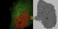
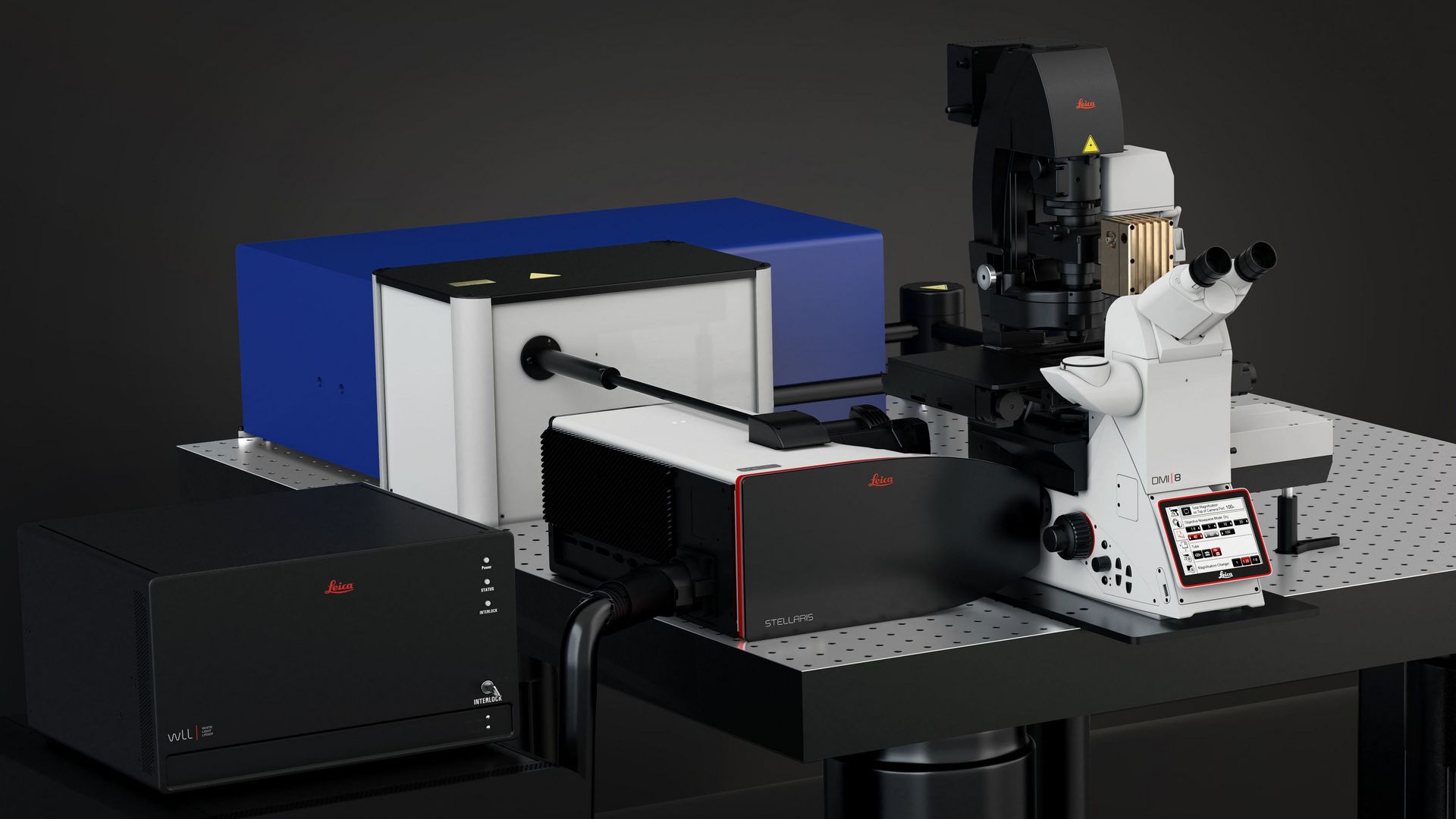
By using advanced Leica imaging solutions that unlock details that were otherwise masked in the image data. An example is out-of-focus blur while imaging deep within cells. Optical sectioning technologies, such as those in the STELLARIS confocal platform or the Viventis Deep Light Sheet imaging system, aid the extraction of insights that reveal fine structures and details, otherwise not visible.

How to accurately quantify cell proliferation/migration?

Leica systems enable precise quantification of migration, proliferation, and differentiation in living cells as well as end point studies. With the support of Aivia machine learning algorithms, biopharma scientists can overcome the manual complexities typically associated with this or similar scientific questions.

How can I enter spatial biology?

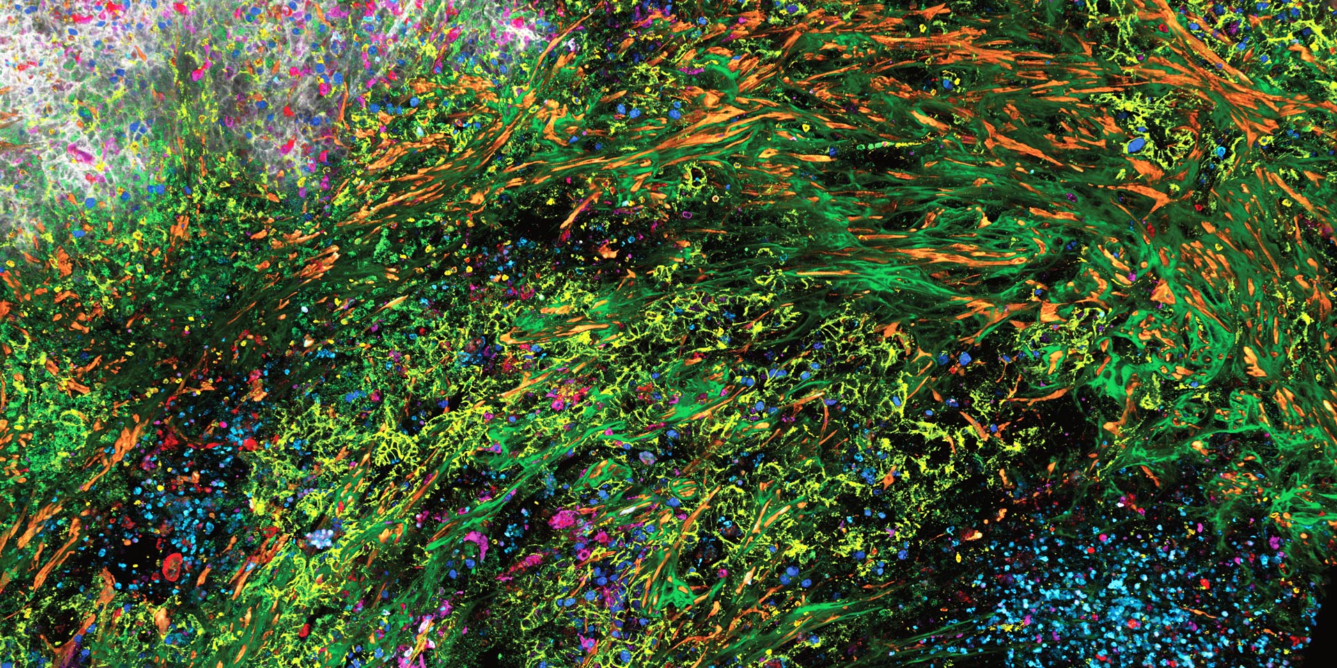
SpectraPlex for STELLARIS grants biopharma researchers access to image complex specimens for spatial biology with 15+ biomarkers in one go, across scales and in 3D. Cell DIVE provides guidelines on antibody usage for iterative multiplexing, with a tested database. Additionally, Aivia enhances spatial biology analysis – integrating it into a standardized, user-friendly protocol.

Can you validate mechanisms with spatial imaging?

Yes, by taking advantage of fluorescence lifetime imaging, super-resolution, multiphoton microscopy, lightsheet and label-free chemical imaging, and 3D high-multiplexing for spatial discoveries in cells and tissues. The STELLARIS confocal platform offers these multimodal capabilities to help validate mechanisms relevant to spatial biology.

What are the advantages for biopharma researchers using Leica advanced microscope solutions?


Data you can trust: CFR-compliant fluorescence imaging for quality control

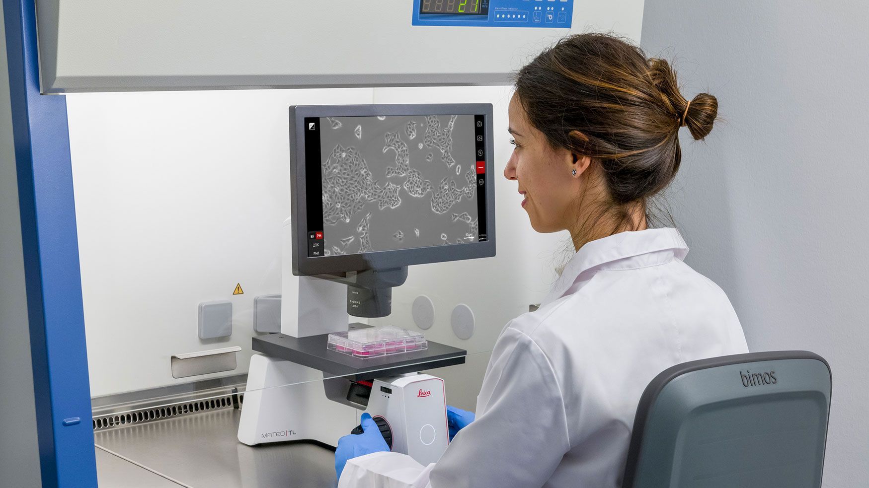
Mateo FL has audit trails and user management functionalities that support FDA 21 CFR Part 11 compliance, making data management easier and more secure for biopharma workflows. 


Label-free molecular imaging for precise analysis

STELLARIS CRS offers label-free molecular imaging, enabling biopharma researchers to overcome the challenge of analyzing complex samples without compromising sample integrity. With fast, non-destructive imaging, it helps researchers tackle molecular analysis in drug discovery, formulation, and disease research.


High-quality data extraction and long-term volumetric imaging of 3D samples

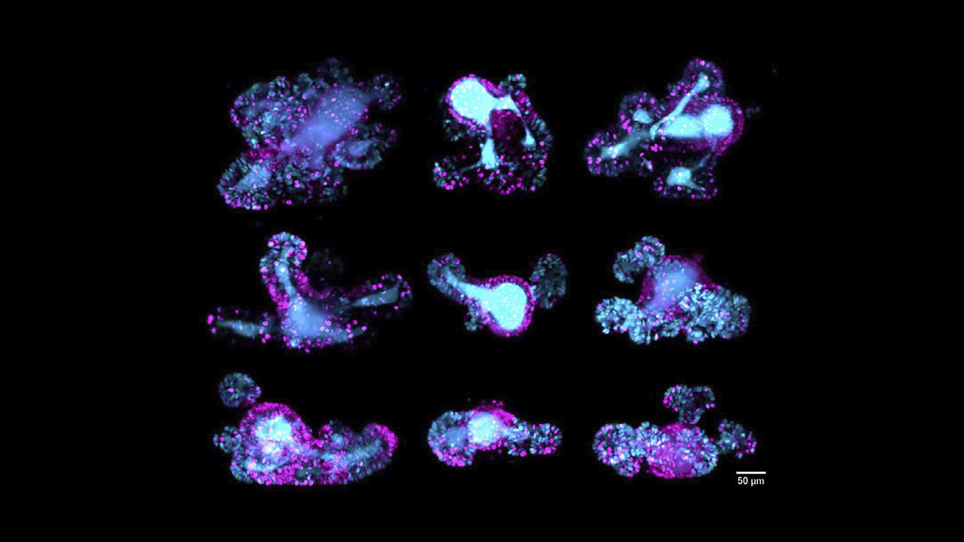
Extract more statistically significant data from challenging 3D samples in less time with high throughput spinning disk confocal. Viventis Deep provides a way to gently visualize up to 2x of the sample volume of multiple organoids in parallel with unprecedented detail over time.

3D high-plex imaging in Cancer Immunology. Overview of a pancreatic tumour section in mouse model, labeled with 15 markers and imaged in one pass using STELLARIS with SpectraPlex. Kunz L., Speziale D., et al., Nat. Methods (2024).

3D high-plex imaging in Cancer Immunology

Overview of a pancreatic tumour section in mouse model, labeled with 15 markers and imaged in one pass using STELLARIS with SpectraPlex. Kunz L., Speziale D., et al., Nat. Methods (2024). 

Characterization of microglial morphology around brain plaques in app/ps1 mice. Imaged using THUNDER Imager Cell Spinning Disk. Shown are both the spinning disk image (left) and the same image after THUNDER Computational Clearing was applied (right). Sample courtesy of Patricia M. Tazo and Aroa S. Maroto, Silvia De Santis’ laboratory, Instituto de Neurociencias CSIC-UMH, Alicante, Spain.

Characterization of microglial morphology around brain plaques in app/ps1 mice

Imaged using THUNDER Imager Cell Spinning Disk. Shown are both the spinning disk image (left) and the same image after THUNDER Computational Clearing was applied (right). Sample courtesy of Patricia M. Tazo and Aroa S. Maroto, Silvia De Santis’ laboratory, Instituto de Neurociencias CSIC-UMH, Alicante, Spain.

67-hour multi-position time-lapse of multiple mouse intestinal organoids in parallel, to follow cell cycle dynamics; scale bar 50 μm. Acquired using Viventis Deep. Maximum intensity projection along the z-axis. Courtesy of Franziska Moos, Liberali group, FMI, Basel, Switzerland.

67-hour multi-position time-lapse of multiple mouse intestinal organoids

67-hour multi-position time-lapse of multiple mouse intestinal organoids in parallel, to follow cell cycle dynamics; scale bar 50 μm. Acquired using Viventis Deep. Maximum intensity projection along the z-axis. Courtesy of Franziska Moos, Liberali group, FMI, Basel, Switzerland.

3D STED imaging of an optically cleared rat kidney section. Nephrin immunolabelled with STAR 635P. Excitation and STED performed with 640 nm and 775 nm, respectively. Image dimensions x,y,z, 83 µm x 83 µm x 40 µm. Volume is color coded for depth. Image courtesy: David Unnersjö Jess, KTH, Stockholm, Sweden.

3D STED imaging of an optically cleared rat kidney section

Nephrin immunolabelled with STAR 635P. Excitation and STED performed with 640 nm and 775 nm, respectively. Image dimensions x,y,z, 83 µm x 83 µm x 40 µm. Volume is color coded for depth. Image courtesy: David Unnersjö Jess, KTH, Stockholm, Sweden.

3D high-plex imaging in Cancer Immunology. Overview of a pancreatic tumour section in mouse model, labeled with 15 markers and imaged in one pass using STELLARIS with SpectraPlex. Kunz L., Speziale D., et al., Nat. Methods (2024).
Characterization of microglial morphology around brain plaques in app/ps1 mice. Imaged using THUNDER Imager Cell Spinning Disk. Shown are both the spinning disk image (left) and the same image after THUNDER Computational Clearing was applied (right). Sample courtesy of Patricia M. Tazo and Aroa S. Maroto, Silvia De Santis’ laboratory, Instituto de Neurociencias CSIC-UMH, Alicante, Spain.
67-hour multi-position time-lapse of multiple mouse intestinal organoids in parallel, to follow cell cycle dynamics; scale bar 50 μm. Acquired using Viventis Deep. Maximum intensity projection along the z-axis. Courtesy of Franziska Moos, Liberali group, FMI, Basel, Switzerland.
3D STED imaging of an optically cleared rat kidney section. Nephrin immunolabelled with STAR 635P. Excitation and STED performed with 640 nm and 775 nm, respectively. Image dimensions x,y,z, 83 µm x 83 µm x 40 µm. Volume is color coded for depth. Image courtesy: David Unnersjö Jess, KTH, Stockholm, Sweden.

Early research: Understanding disease mechanisms

Early-stage biopharma research focuses on uncovering the cellular and molecular mechanisms of diseases. High-resolution imaging is essential to visualize biological structures and interactions in their native context. 

Leica advanced confocal, super-resolution, and widefield microscopy systems enable precise imaging of cells. They support live-cell imaging and time-lapse studies, providing critical insights into cellular behavior and disease progression. With intuitive Leica software and AI-enhanced image analysis, researchers can accelerate data interpretation and hypothesis generation.

3D 15-plex imaging for phenotyping in immune response. Image acquired using STELLARIS with SpectraPlex. Roberti J., Hecht F., Gai E., Straka T., Holzmeister S., Steinmetz I., Wong H., Alvarez L. Nat. Methods (2024).

Drug discovery: Target identification and validation

In drug discovery, identifying and validating biological targets is crucial. Visualizing protein expression, biomarker distribution, and cellular responses play a critical role in this process.Researchers need cutting-edge imaging tools to support immunofluorescence, and protein localization studies. 

Leica confocal and super-resolution microscopes deliver the clarity and speed needed to screen and analyze large sample sets. Automation capabilities streamline acquisition and analysis, enabling reproducibility and reducing human error.

Drug manufacturing & quality control: Ensuring consistency and compliance

In manufacturing and quality control (QC) environments, maintaining product integrity, sterility, and regulatory compliance is non-negotiable. Visual inspection plays a key role in validating each step.

Mateo FL Digital Fluorescence Microscope helps to boost advanced cell culture research with its multi-modal fluorescence and transmitted light capabilities, automated analysis tools and secure data tracking. It has built-in audit trails and user management functionalities that support FDA 21 CFR Part 11 compliance, for easier and secure data management.

Brain organoids labeled with lamin (green) and tubulin (magenta), acquired using Viventis Deep Dual View Light Sheet Microscope. Courtesy of Akanksha Jain, Treutlein Lab ETH-DBSSE Basel (Switzerland).

Drug development: Preclinical and translational research

As drug candidates move into development, rigorous testing and deeper biological insights become essential. Imaging helps researchers to evaluate efficacy, safety, and the mechanism of action.

The THUNDER Imager Cell with Spinning Disk from Leica Microsystems provides researchers with high quality data and clear details from 3D samples for a deeper and more detailed view which can help advance their research.

Frequently asked questions in biopharma

Show answer Why leverage live dead assays during drug discovery and development?

Patient safety is the highest priority. Therefore, knowledge about lethal dosages is invaluable and required by regulatory agencies before considering clinical trials. Live and dead assays can be utilized to determine the limits. For efficient acquisition of an adequate quantity of data, Mica is an optimal solution.

Show answer How can biopharma research be improved with laser microdissection?

By providing precise tissue isolation, enabling high-quality genomic and proteomic analysis. Laser microdissection helps biopharma researchers target specific cells or tissue regions, facilitating more accurate and contamination-free studies in drug development.

Show answer Why use microscopy for biopharma research concerning drug discovery and development?

Microscopy can help with gaining detailed spatial knowledge of cellular responses to compounds and drug candidates. During the drug discovery phase, i.e., quality control and in-depth analysis of response pathways for both living cell culture models and tissues, there are a range of Leica products that meet different discovery and development needs.

Related Articles

Read our latest articles about imaging solutions for biopharma

The knowledge portal of Leica Microsystems offers scientific research and teaching material on the subjects of microscopy. The content is designed to support beginners, experienced practitioners and scientists alike in their everyday work and experiments.

More Articles
Brain organoid labeled with lamin (green) 
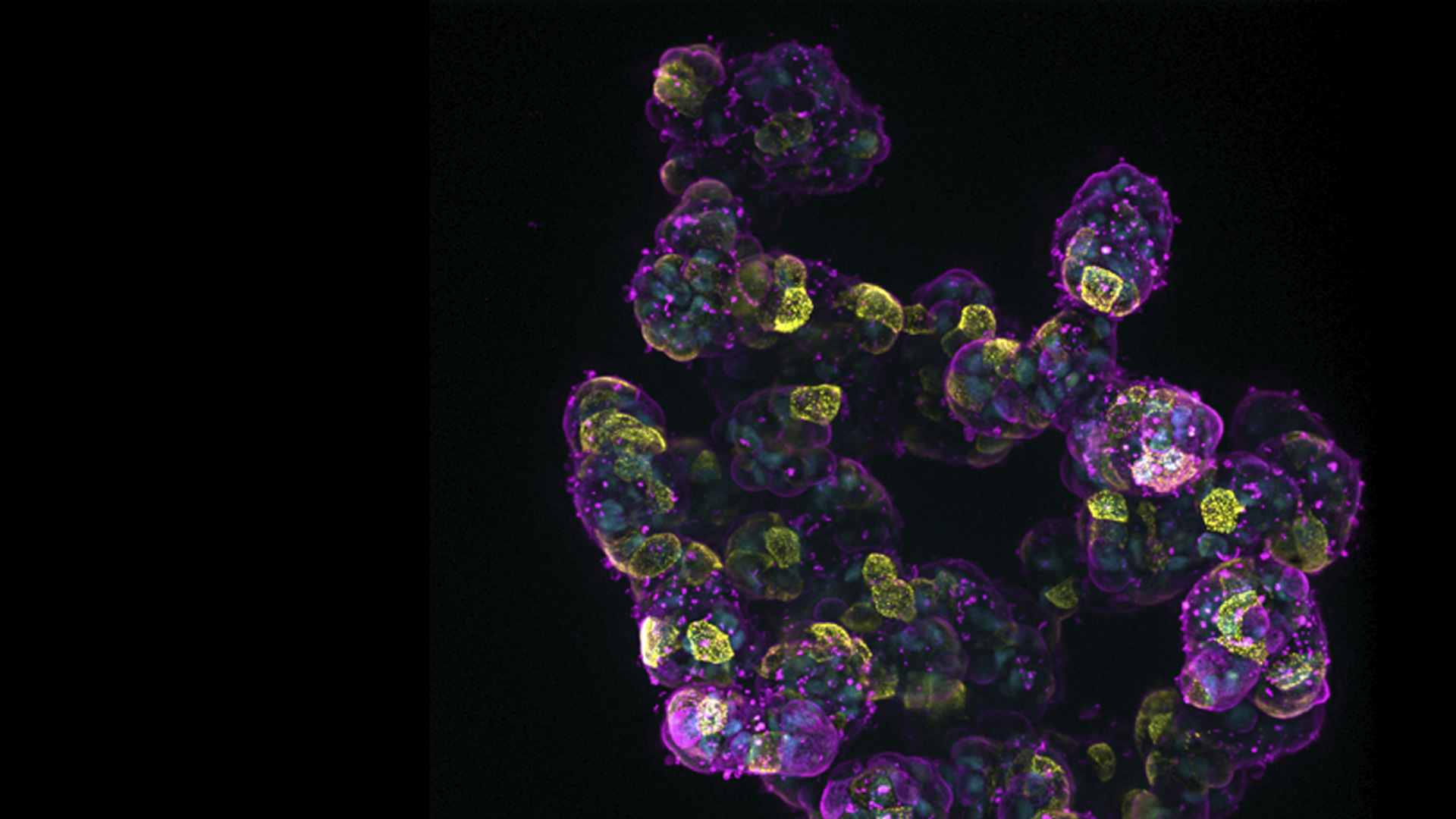
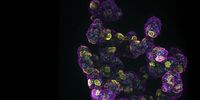
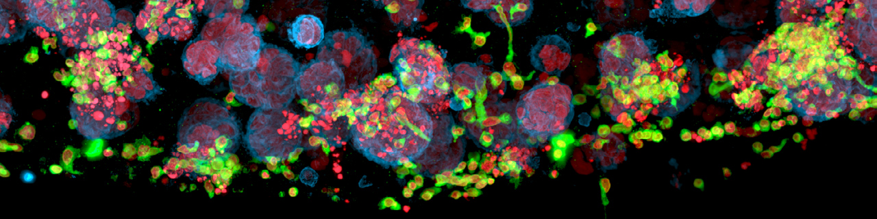
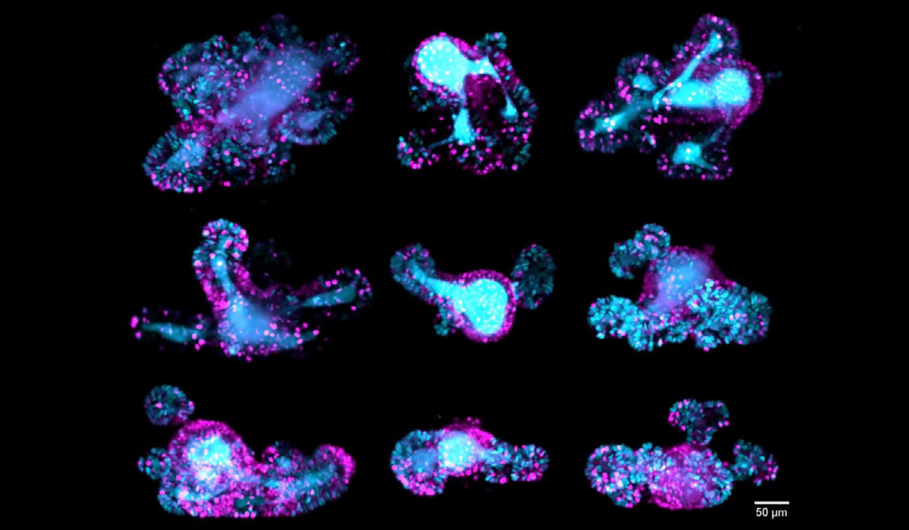
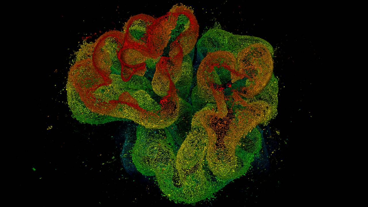
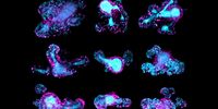
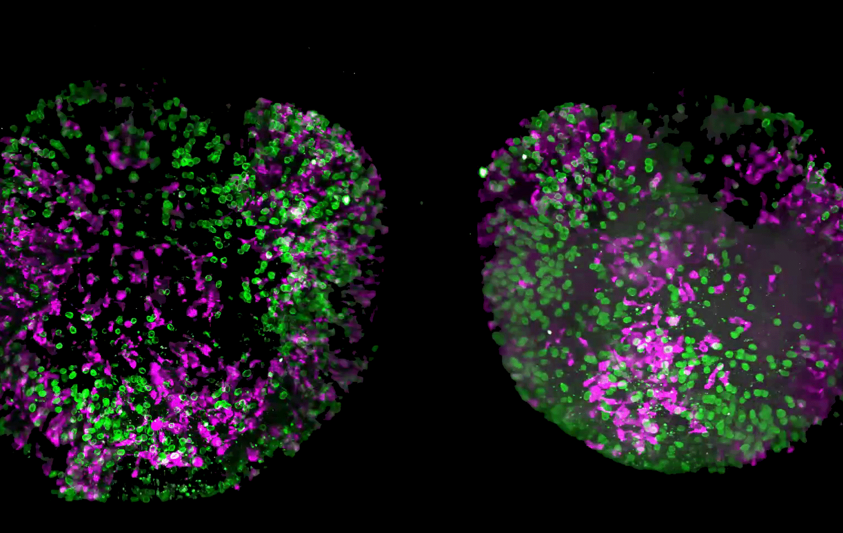
and tubulin (magenta), acquired using Viventis Deep. Courtesy of Akanksha Jain, Treutlein Lab ETH-DBSSE Basel (Switzerland).

Faster & Deeper Insights into Organoid and Spheroid Models

Gain deeper, more translatable, insights into organoid and spheroid models for drug discovery and disease research by overcoming key imaging challenges. In this eBook, explore advanced microscopy…
Pancreatic Ductal Adenocarcinoma with 11 Apoptosis biomarkers shown – BAK, BAX, BCL2, BCLXL, Caspase9, CIAP1, NaKATPase, PCK26, SMAC, Vimentin, and XIAP.

Transforming Research with Spatial Proteomics Workflows

Spatial Proteomics, Nature Methods 2024 Method of the Year, is driving research advancements in cancer, immunology, and beyond. By combining positional data with high throughput imaging of proteins in…
Digital microscopy simplifies documenting cell-culture results electronically while following 21 CFR part 11 guidelines for biopharma.

Introduction to 21 CFR Part 11 for Electronic Records of Cell Culture

This article provides an introduction to the recommendations of 21 CFR Part 11 from the FDA, specifically focusing on the audit trail and user management in the context of cell-culture laboratories.…
Automated Laser Microdissection for Proteome Analysis

Deep Visual Proteomics Provides Precise Spatial Proteomic Information

Despite the availability of imaging methods and mass spectroscopy for spatial proteomics, a key challenge that remains is correlating images with single-cell resolution to protein-abundance…
AI-based transfection analysis (left) 
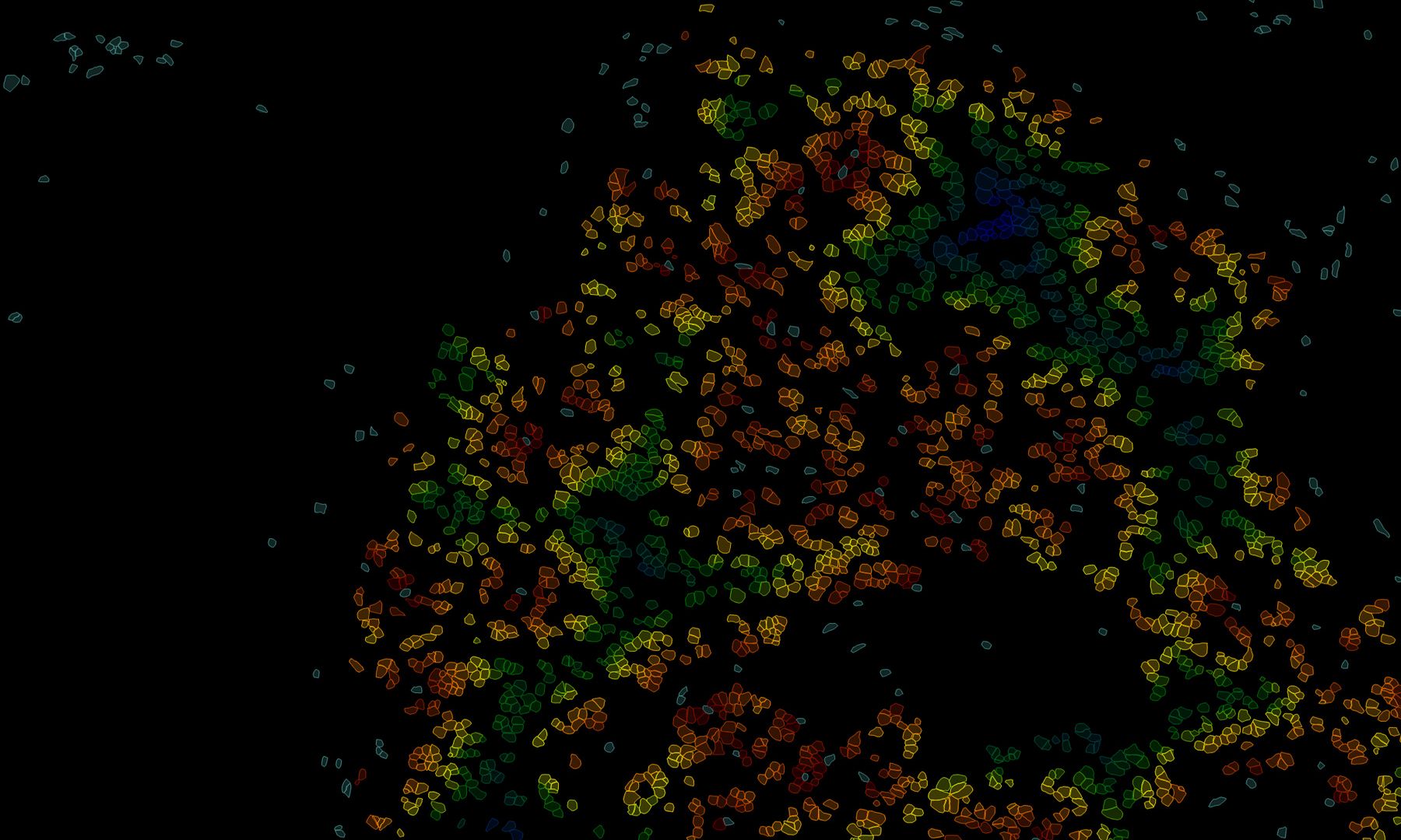
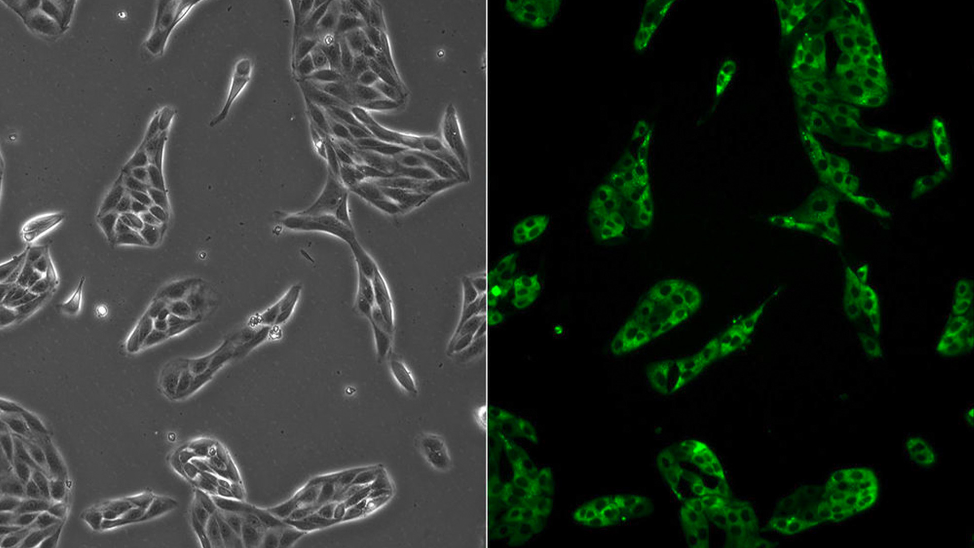
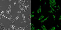
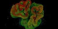
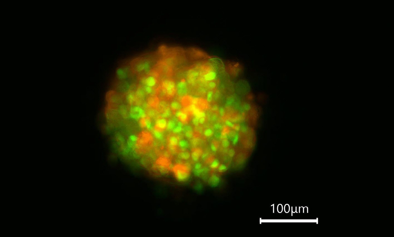
of U2OS cells which were transfected with a fluorescently labelled protein. A fluorescence image of the cells (right) is also shown. The analysis and imaging were performed with Mateo FL.

Leveraging AI for Efficient Analysis of Cell Transfection

This article explores the pivotal role of artificial intelligence (AI) in optimizing transfection efficiency measurements within the context of 2D cell culture studies. Precise and reliable…
Living HeLa cells stained with WGA-488 (yellow)
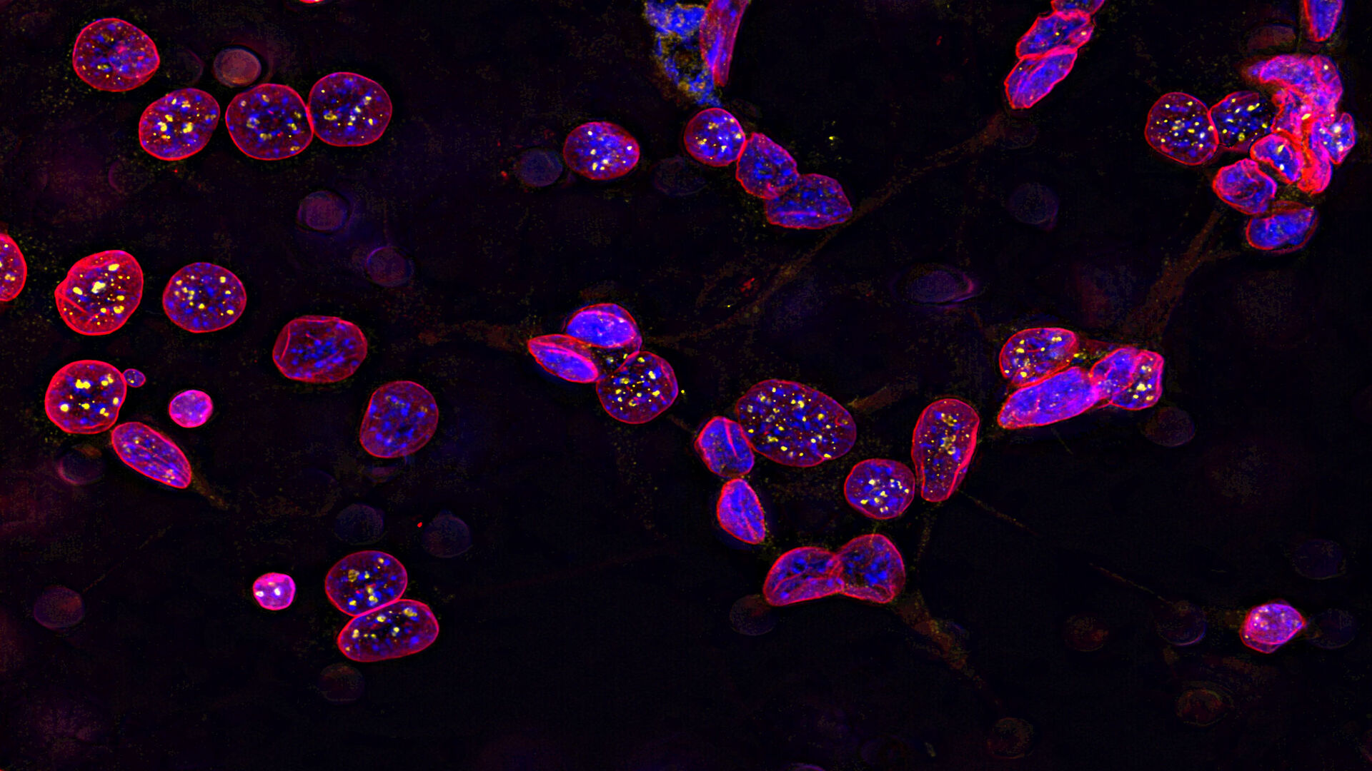
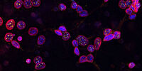
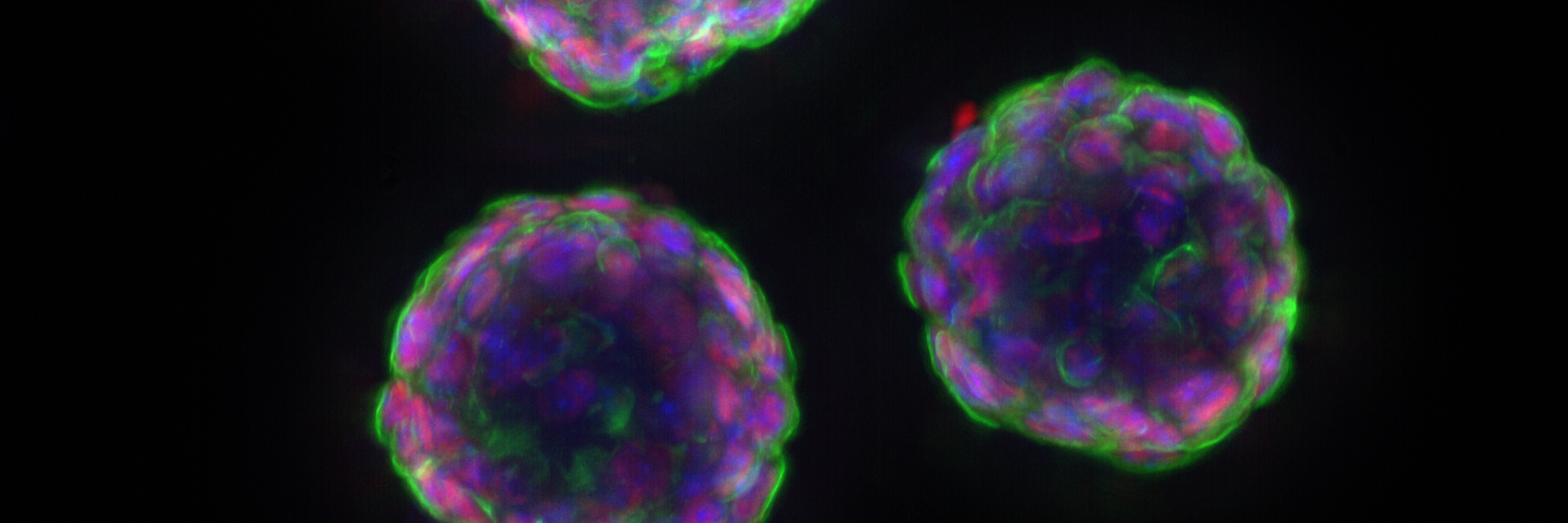
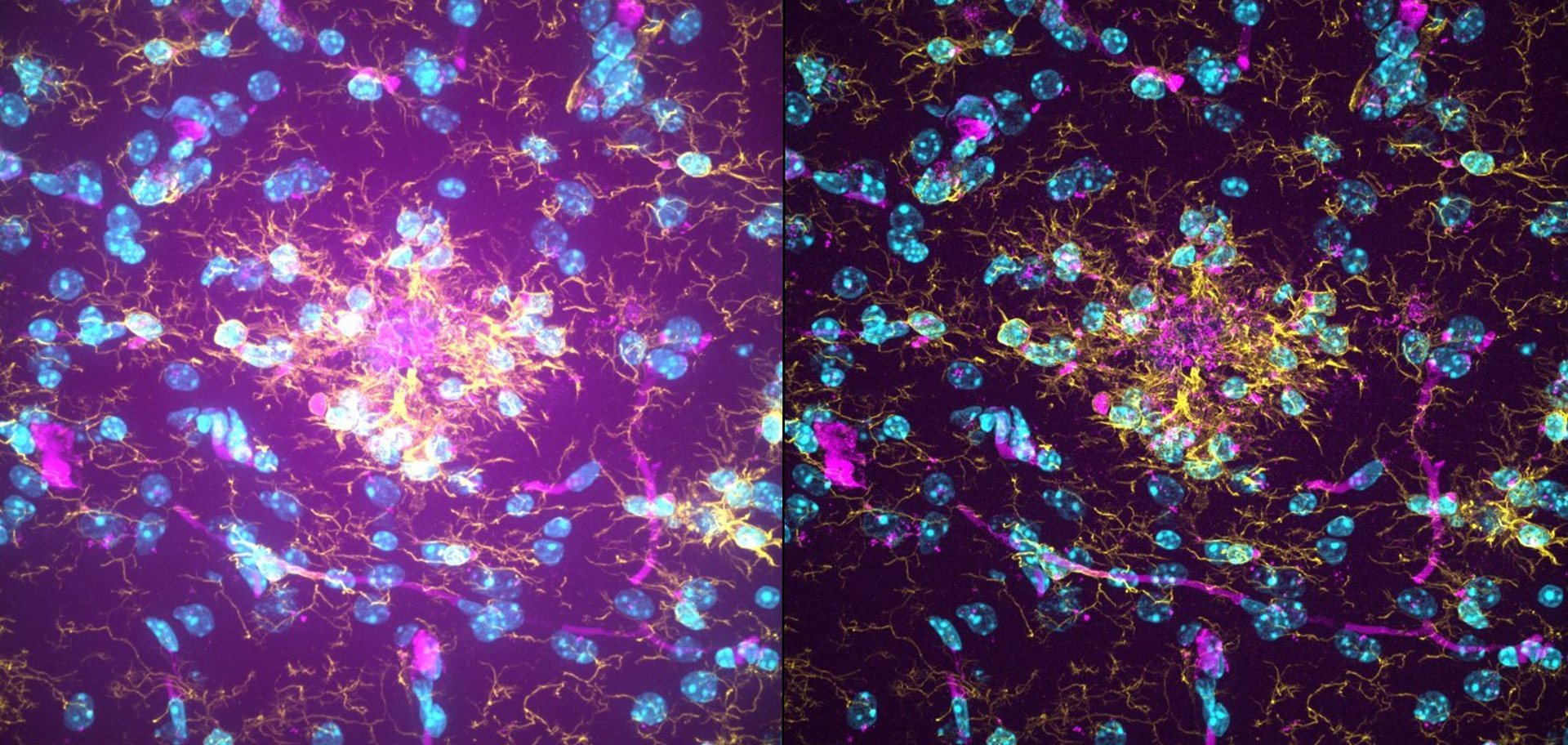
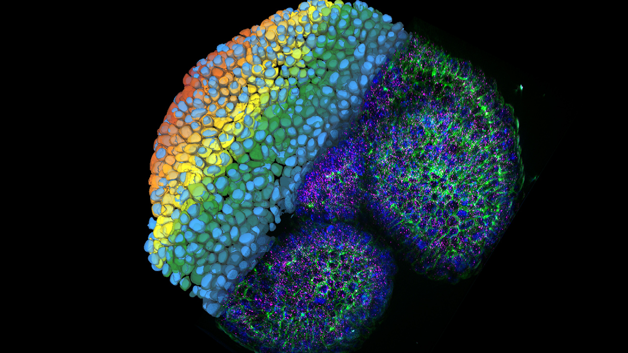
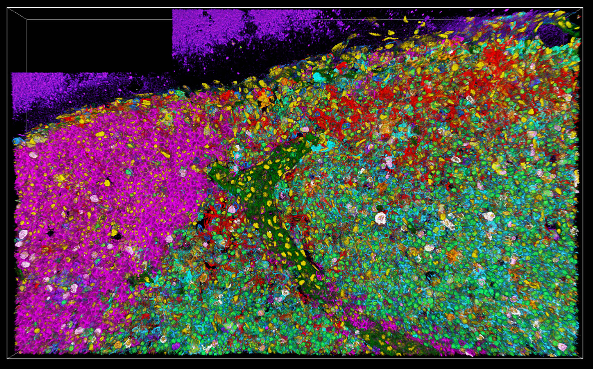
, SPY-Actin (cyan), and SiR-Tubulin (magenta). Instant Computational Clearing (ICC) was applied.

How to Perform Dynamic Multicolor Time-Lapse Imaging

Live-cell imaging sheds light on diverse cellular events. As many of these events have fast dynamics, the microscope imaging system must be fast enough to record every detail. One major advantage of…
Virally labeled neurons (red) and astrocytes (green) in a cortical spheroid derived from human induced pluripotent stem cells. THUNDER Model Organism Imager with a 2x 0.15 NA objective at 3.4x zoom was used to produce this 425 µm Z-stack (26 positions), which is presented here as an Extended Depth of Field (EDoF) projection.  Images courtesy of Dr. Fikri Birey  from the Dr. Sergiu Pasca laboratory at Stanford University, 3165 Porter Dr., Palo Alto, CA

Download The Guide to Live Cell Imaging

In life science research, live cell imaging is an indispensable tool to visualize cells in a state as in vivo as possible. This E-book reviews a wide range of important considerations to take to…
14: 7-day old 3D cell culture of MDCK cells stably transfected with; Mx1-GFP (green), SPY555-Actin (yellow) and WGA-AlexaTM 647 (magenta), and imaged using a THUNDER Imager Cell Spinning Disk. Shown are both the spinning disk image (left) and the same image after THUNDER Computational Clearing was applied (right); scale bar 50 μm.

Organoids and 3D Cell Culture

One of the most exciting recent advancements in life science research is the development of 3D cell culture systems, such as organoids, spheroids, or organ-on-a-chip models. A 3D cell culture is an artificial environment in which cells are able to grow and interact with their surroundings in all 3 dimensions. These conditions are similar to how they would be in vivo.

Read More

Example of the heat map of the nearest neighbor between Cluster 3 to 12 by Aivia Software

Cellular Analysis

Ranging from functional to correlative imaging, the versatility of Leica microscopy solutions ensures that researchers have the right tools tailored to their specific scientific applications and experimental goals.

Read More

Image of C2C12 cells: The cells are stained with lamin B (magenta) which indicates nuclear structure, Hoechst (blue) indicating DNA, and γH2AX (yellow) indicating damage to DNA. Cells were imaged using a THUNDER Imager 3D Live Cell with a 63X/1.4 oil immersion objective.

Cell Biology Research

Choosing the right microscopy method is critical if you want to maximize your research output and obtain the highest quality data. Leica Microsystems offers various solutions to improve your cell biology research.

Read More

Mammalian cells expressing H2B-mCherry and alphaTubulin-mEGFP to visualize DNA and microtubules respectively. Left: Widefield imaging using THUNDER Imager. Right: Sample after correlated EM imaging using Leica Microsystems Coral Life workflow. Cells were cryo-immobilized by high-pressure freezing 60 min after the onset of cytokinesis. The intercellular bridge was imaged under TEM after freeze substitution and resin infiltration.

Correlative Light and Electron Microscopy Solutions

CLEM solutions from Leica Microsystems ensure sample viability and quality checks. Users can take advantage of these solutions to identify directly the right cell at the right time, obtain high-resolution cryo-confocal data, or put the fluorescence information into the ultrastructural context.

Read More

Cell Culture

Growing cells under lab conditions is the base for scientists working in fields of cell or developmental biology, cancer research, or any kind of life science and pharma research. Find out how Leica cell culture solutions PAULA, DMi1 and DM IL help to culture animal cells in the lab.

Read More

14: 7-day old 3D cell culture of MDCK cells stably transfected with; Mx1-GFP (green), SPY555-Actin (yellow) and WGA-AlexaTM 647 (magenta), and imaged using a THUNDER Imager Cell Spinning Disk. Shown are both the spinning disk image (left) and the same image after THUNDER Computational Clearing was applied (right); scale bar 50 μm.
Example of the heat map of the nearest neighbor between Cluster 3 to 12 by Aivia Software
Image of C2C12 cells: The cells are stained with lamin B (magenta) which indicates nuclear structure, Hoechst (blue) indicating DNA, and γH2AX (yellow) indicating damage to DNA. Cells were imaged using a THUNDER Imager 3D Live Cell with a 63X/1.4 oil immersion objective.
Mammalian cells expressing H2B-mCherry and alphaTubulin-mEGFP to visualize DNA and microtubules respectively. Left: Widefield imaging using THUNDER Imager. Right: Sample after correlated EM imaging using Leica Microsystems Coral Life workflow. Cells were cryo-immobilized by high-pressure freezing 60 min after the onset of cytokinesis. The intercellular bridge was imaged under TEM after freeze substitution and resin infiltration.
Scroll to top