Introduction
Neuroscience is a multidisciplinary field involving the study of the structure and function of the nervous system. The purpose is to understand the development of cognitive and behavioral processes as well as understand and find therapies for disorders, such as Alzheimer’s or Parkinson’s disease.
The use of microscopy techniques is critical to visualize the nervous system at cellular and subcellular levels and view any molecular changes within context. Recent developments in deep tissue imaging have provided further insights into neuronal function. Emerging technologies like genetic cell labeling and optogenetics complement these developments.
Imaging challenges for neuroscience research
Research of the nervous system often requires the combination of high resolution, deep imaging and visualization of large sections. You also require flexibility to image different types of samples, such as live cells, tissues, organoids, and model organisms.
The study of fast dynamic processes, such as cell transport or synaptic remodeling, require high-speed microscopy. One of the main challenges of high-speed microscopy is acquiring high-resolution images while avoiding fluorescence saturation.
Neuroscience research often involves wide-area and volumetric imaging. The need to reduce fluorescence scattering and the background signal can make acquiring images with high contrast and resolution difficult, which is particularly critical when examining neuronal architecture in dense tissues like brain sections.


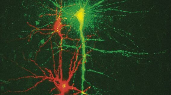
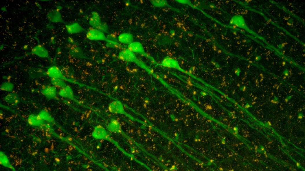
Cultured cortical neurons. Z-stack of 59 planes (thickness: 21µm). Sample courtesy of FAN GmbH, Magdeburg, Germany.
Related articles
What are the Challenges in Neuroscience Microscopy?

Neuroscience Images
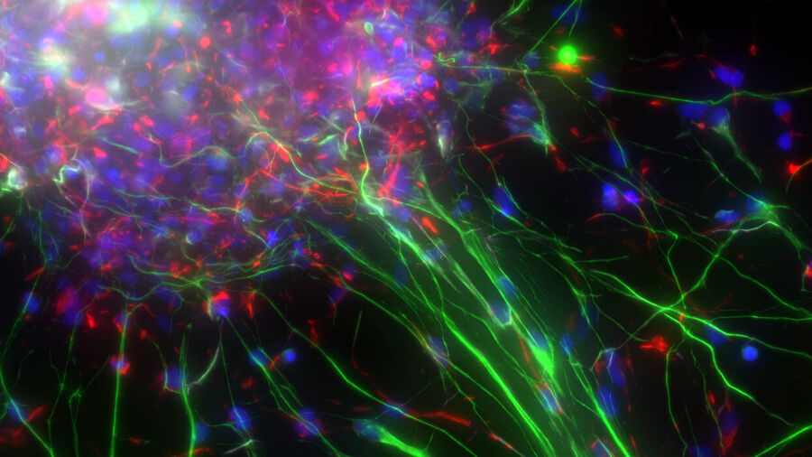
 and astrocytes (green) in a cortical spheroid derived from human induced pluripotent stem cells.")
How do Cells Talk to Each Other During Neurodevelopment?

Imaging Organoid Models to Investigate Brain Health
 acquired using THUNDER Imager Live Cell. Image courtesy of Janina Kaspar and Irene Santisteban, Schäfer Lab, TUM.")
Microscopy methods for neuroscience research
The study of the nervous system typically relies on confocal microscopy for high resolution imaging of events and structures. For deeper in vivo imaging, multiphoton microscopy is used, as its capacity to use near-infrared excitation reduces light scattering, enabling deep imaging with minimal invasiveness. Lightsheet microscopy is also preferred for light-sensitive or 3D samples. It reduces phototoxicity while providing intrinsic optical sectioning and 3D imaging.
- Optogenetics is a technique that involves controlling neural activity using light and enables the study of specific neuronal networks and cell signaling. It requires the expression of light-sensitive proteins in the neuronal cell membrane. Exploring events at the nanoscale using optogenetics in combination with timed millisecond precision vitrification is a promising technology to study specific time points within a dynamic process.
- Electrophysiology is the study of the electrical properties of tissues and cells and includes the study of the electrical properties of neurons. The function of nerve and muscle cells relies on ionic currents flowing through ion channels. One way to investigate ion channels is to use patch clamping. This method allows investigation of ion channels in detail and recording of the electric activity of different types of cells, mainly excitable cells like neurons.


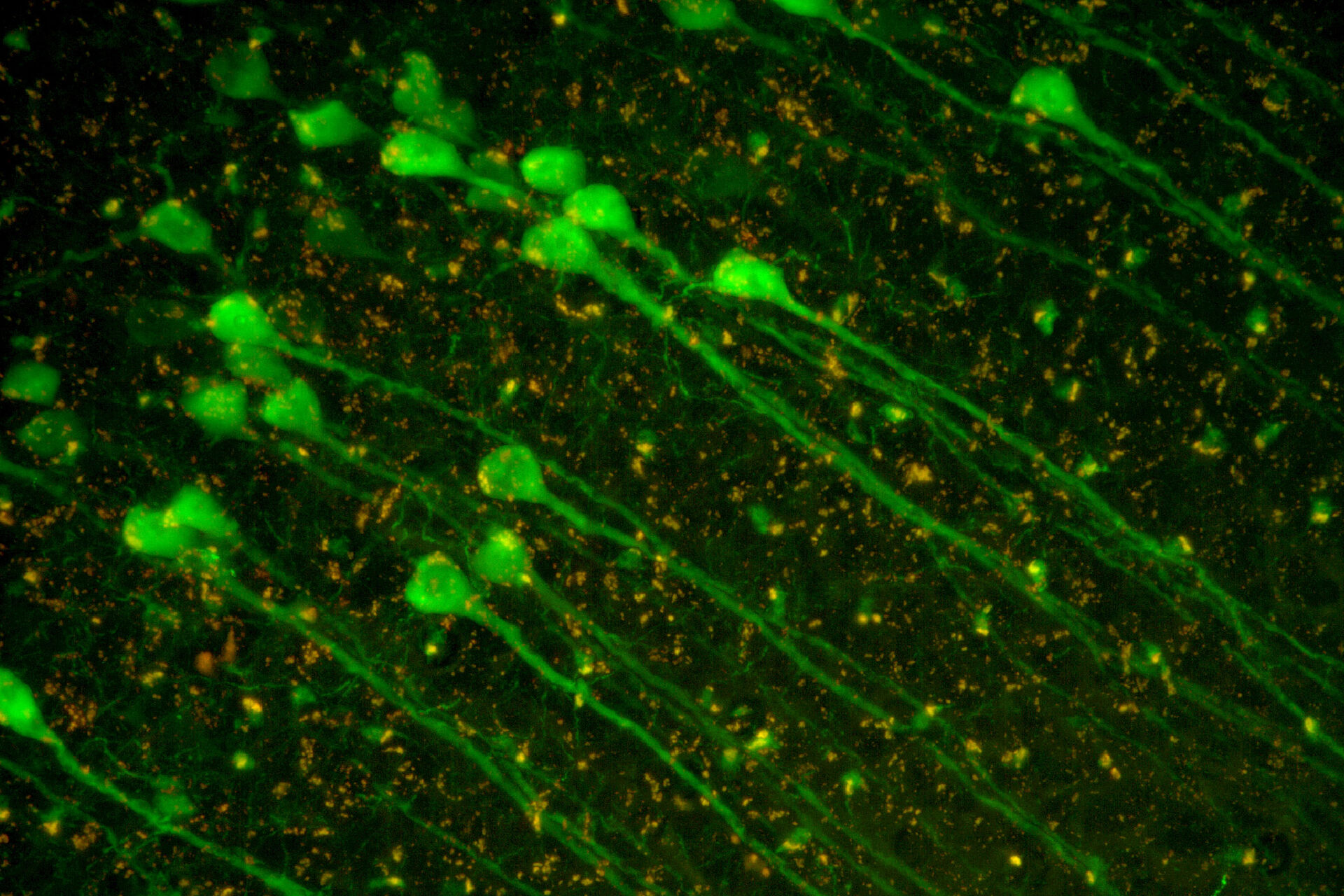
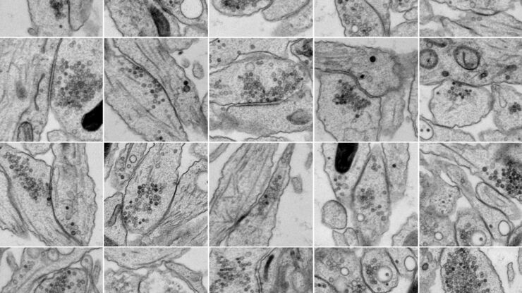
. Image courtesy of Prof. Hui Guo, School of Life Sciences, Central South University, China")
.")