Neuro-Oncology Surgery Solutions
Suspected Grade III and IV gliomas are aggressive brain tumors needing precise surgery. At Leica Microsystems, we have developed two neuro-oncological solutions: FL400 and GLOW400 3D AR fluorescence modules. Both solutions, in conjunction with 5-ALA, enable better visualization.
Contact a local specialist for expert advice on the right Neuro-Oncology microscope for your needs and budget.

Why is clear visualization crucial during glioma surgeries?
High-grade gliomas can infiltrate critical brain regions, making clear visualization essential for surgery. Leica Microsystems' surgical microscopes provide precision optics and integrate image-guided surgery (IGS) systems for advanced neuronavigation, offering surgeons detailed visual information. This integration enhances decision-making and ensures precise, informed surgical procedures.

How can fluorescence-guided surgery improve visualization in tumor resection?
Balancing tumor resection with preserving neurological function is crucial. Fluorescence-guided surgery, using 5-ALA, helps visualize tumors by highlighting them under blue light. Our FL400 and GLOW400 3D AR modules enhance visualization of suspected Glioma III and IV tumors, aiding neurosurgeons in achieving optimal patient outcomes.

How does fluorescence-guided surgery support tumor resection and patient prognosis?
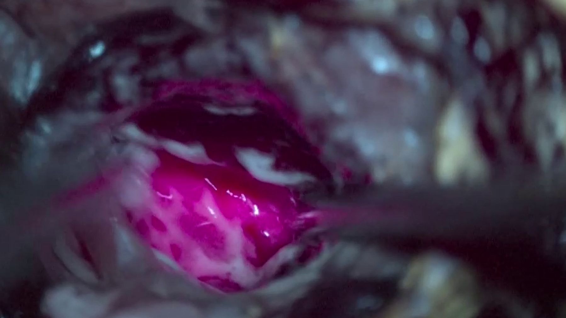
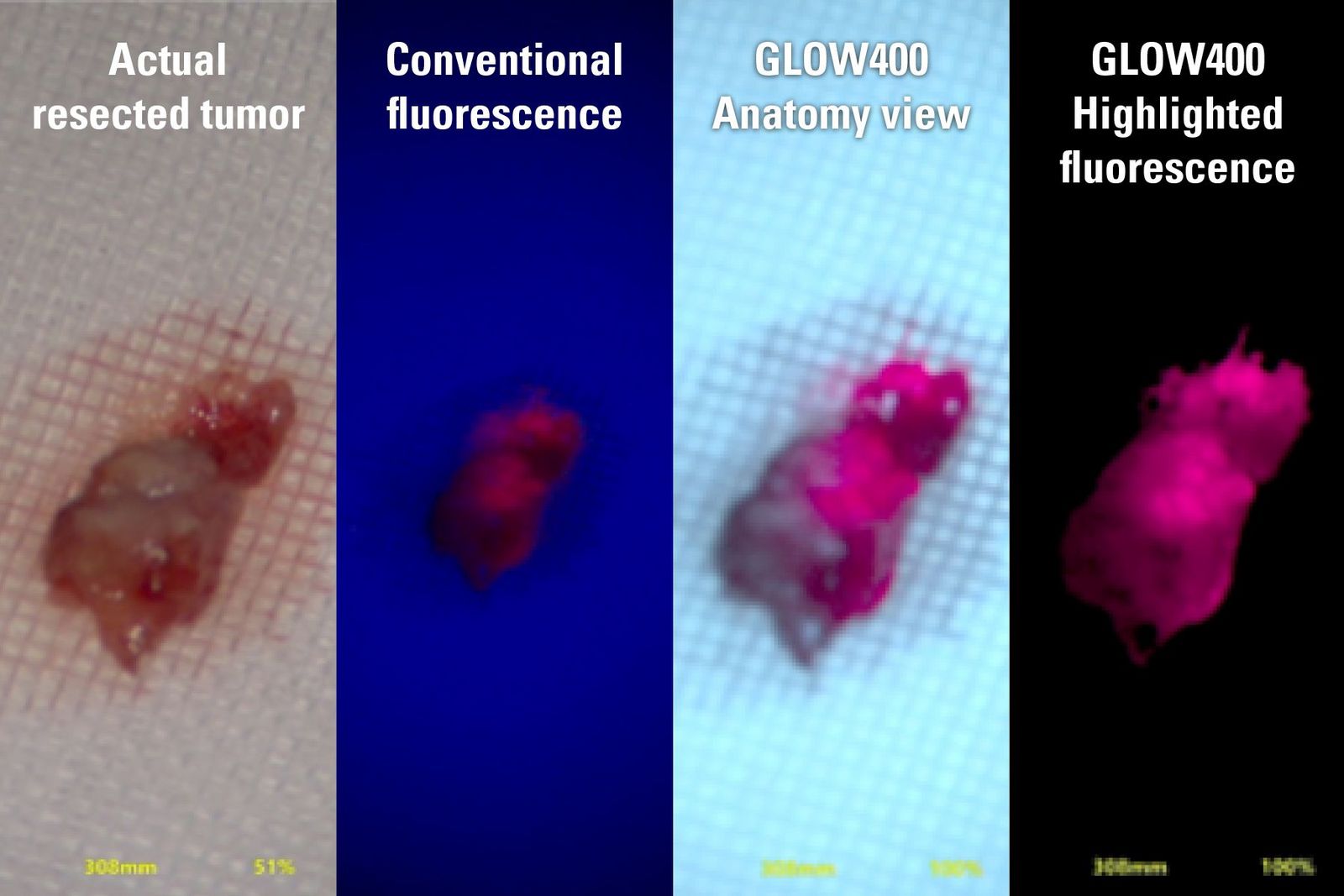
Fluorescence-guided surgery can help maximize malignant tumor removal. The FL400 module highlights the tumor in pink under classic blue light visualization in the oculars. GLOW400 3D AR uses multispectral imaging technology, providing an enhanced anatomical visualization augmented with the fluorescent marked tumor, allowing the surgeon to better see critical structures during resection.

How does fluorescence-guided surgery with 5-ALA improve surgical workflow?
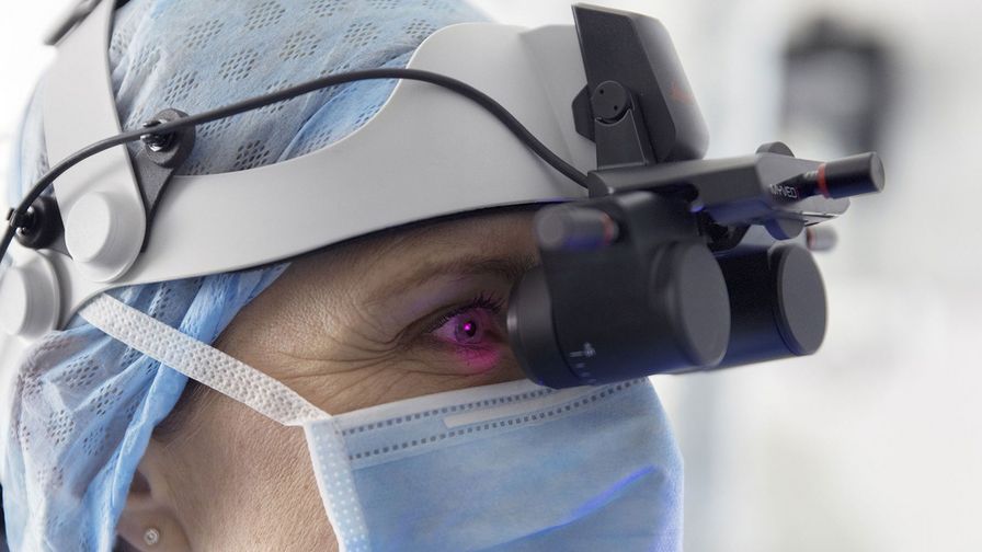
Studies show fluorescence-guided surgery with 5-ALA can improve resection outcomes. The GLOW400 application enhances workflow by eliminating the need of having to switch between different microscope views, reducing fatigue and interruptions. Surgeons can access real-time anatomical information in 2D for observation and conduct 3D heads-up surgery via large monitor. With the MyVeo headset, GLOW400 can be seen directly in front of the eyes for an optimal intraoperative workflow.
What makes Leica surgical microscopes an excellent choice for neuro-oncology surgery?
Enhanced optical visualization
FusionOptics by Leica Microsystems boosts stereoscopic microscopy by utilizing two different optical pathways: one with a high resolution and one with a greater depth of field. The human brain merges this information into a single, clear 3D image. The result is an image with improved depth of field and resolution for precise surgical visualization.
Augmented Reality Fluorescence Application
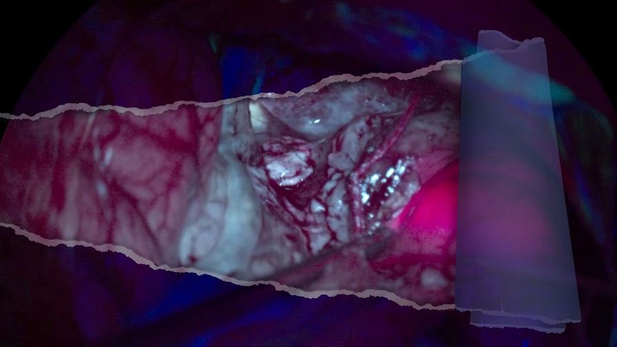
The GLOW400 Anatomy view merges fluorescent and non-fluorescent tissue information in real-time, enhancing tumor visualization. During resection, the HiFluo view improves detection of low-intensity fluorescence in suspected Grade III and IV gliomas.
Boosted collaboration & enhanced teaching
The MyVeo headset for the ARveo 8 microscope displays essential clinical data directly in front of your eyes, enhancing focus and comfort. Up to three users can experience real-time surgery simultaneously, making it a powerful tool for teaching and learning.

Unmatched Service
We offer different Service plans to fit your needs designed to provide you with full coverage for systems that are critical to your workflow. Reducing operational risk and optimizing performance with minimal disruption is of utmost importance. Learn more here.
How to achieve brain tissue resection with GLOW400 AR
Clinical case by Prof. Kondo
Read our latest clinical case on brain tumor surgery using GLOW400 3D AR fluorescence, by Prof. Akihide Kondo, Chief Professor in the Department of Neurosurgery at Juntendo University Hospital, Tokyo, Japan. Learn how advanced AR technology can help enhance tumor resection precision.
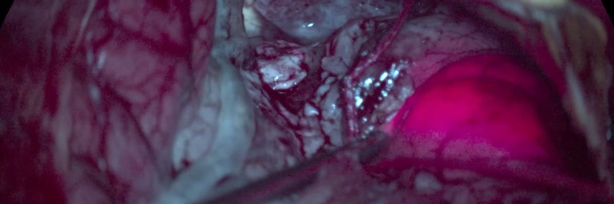
See clearer anatomical details surrounding the fluorescent-marked tumor
Suspected Grade III and IV gliomas are aggressive brain tumors requiring precise surgery. Leica's FL400 and GLOW400 3D AR fluorescence modules aid in tumor visualization.

During suspected grade III and IV glioma surgeries, GLOW400 allows to see clearer anatomical details surrounding the fluorescent-marked tumor through the Anatomy View. Image courtesy of Tim Jacquesson, MD, PhD Hospices Civils de Lyon, France.
Related Articles

How to Achieve Brain Tissue Resection with GLOW400 AR
 set-up.")
Augmented Reality: Transforming Neurosurgical Procedures

3D, AR & VR for Teaching in Neurosurgery

Surgical Management of High-Grade Gliomas

Augmented Reality Assisted Navigation in Neuro-Oncological Surgery
Frequently asked questions Neuro-Oncology surgery solutions
The aim of brain tumor surgery is to remove as much of the tumoral tissue as possible, alleviating symptoms caused by pressure on brain structures. Surgery also allows for a biopsy to determine the tumor type and guide further treatment. Advanced techniques like craniotomy and stereotactic radiosurgery are used based on the tumor's location and size. Pre-surgical planning with imaging and functional mapping helps minimize risks to critical brain functions. In certain cases, surgery is followed by radiation or chemotherapy to target remaining tumor cells.
High-grade gliomas, such as glioblastomas, require precise surgical tumor removal. Fluorescence imaging helps mark the tumor and delineate tumor margins for optimal surgical outcome and extent of resection.
When selecting a neuro-oncological surgery microscope, high-resolution optics are essential for clear and detailed visualization of brain tissues. Fluorescence imaging is crucial as it helps differentiate between healthy and tumor tissues, enhancing surgical visualization. Additionally, an ergonomic design is important for comfort during long surgeries. Augmented reality (AR) can overlay digital information on the surgical field, and image-guided surgery (IGS) integration assists in precise navigation of tumor areas. These features collectively improve precision, and safety in neuro-oncological surgeries.
