
Spécialités médicales
Spécialités médicales
Explorez une collection complète de ressources scientifiques et cliniques conçues pour les professionnels de la santé, notamment des points de vue de pairs, des études de cas cliniques et des symposiums. Conçue pour les neurochirurgiens, les ophtalmologues et les spécialistes en chirurgie plastique et reconstructive, en ORL et en dentisterie. Cette collection met en lumière les dernières avancées en matière de microscopie chirurgicale. Découvrez comment les technologies chirurgicales de pointe, telles que la fluorescence AR, la visualisation 3D et l'imagerie OCT peropératoire, permettent de prendre des décisions en toute confiance et d'être précis dans les chirurgies complexes.

Live Cell Imaging Gallery
Live cell microscopy techniques are fundamental to get a better understanding of cellular and molecular function. Today, widefield microscopy is the most common technique used to visualize cell…

Tissue Image Gallery
Visual analysis of animal and human tissues is critical to understand complex diseases such as cancer or neurodegeneration. From basic immunohistochemistry to intravital imaging, confocal microscopy…
 and astrocytes (green) in a cortical spheroid derived from human induced pluripotent stem cells.")
Neuroscience Images
Neuroscience commonly uses microscopy to study the nervous system’s function and understand neurodegenerative diseases.

Multicolor Image Gallery
Fluorescence multicolor microscopy, which is one aspect of multiplex imaging, allows for the observation and analysis of multiple elements within the same sample – each tagged with a different…
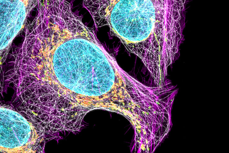
Cell Biology Image Gallery
Cell biology studies the structure, function and behavior of cells, including cell metabolism, cell cycle, and cell signaling. Fluorescence microscopes are an integral part of a cell biologist…

Kinetochore Assembly during Mitosis with TauSTED on 3D
Three-dimensional organization of the mitotic spindle together with the distribution of CENP-C and BUB1 based on TauSTED with multiple STED lines (592, 660 and 775 nm) can provide insights…

How to Quantify Changes in the Metabolic Status of Single Cells
Metabolic imaging based on fluorescence lifetime provides insights into the metabolic dynamics of cells, but its use has been limited as expertise in advanced microscopy techniques was needed.
Now,…

Regulators of Actin Cytoskeletal Regulation and Cell Migration in Human NK Cells
Dr. Mace will describe new advances in our understanding of the regulation of human NK cell actin cytoskeletal remodeling in cell migration and immune synapse formation derived from confocal and…

Benefits of TauContrast to Image Complex Samples
In this interview, Dr. Timo Zimmermann talks about his experience with the application of TauSense tools and their potential for the investigation of demanding samples such as thick samples or…
