
Spécialités médicales
Spécialités médicales
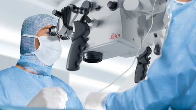
Explorez une collection complète de ressources scientifiques et cliniques conçues pour les professionnels de la santé, notamment des points de vue de pairs, des études de cas cliniques et des symposiums. Conçue pour les neurochirurgiens, les ophtalmologues et les spécialistes en chirurgie plastique et reconstructive, en ORL et en dentisterie. Cette collection met en lumière les dernières avancées en matière de microscopie chirurgicale. Découvrez comment les technologies chirurgicales de pointe, telles que la fluorescence AR, la visualisation 3D et l'imagerie OCT peropératoire, permettent de prendre des décisions en toute confiance et d'être précis dans les chirurgies complexes.
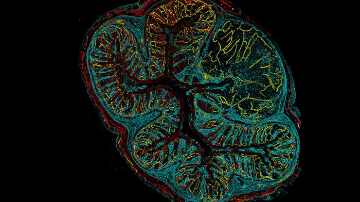
Visualizing Life in 3D at High Speed with Viventis SCAPE
Discover how oblique plane microscopy (OPM) enables light sheet imaging through a single objective, combining illumination and detection in a geometry that is compatible with conventional sample…
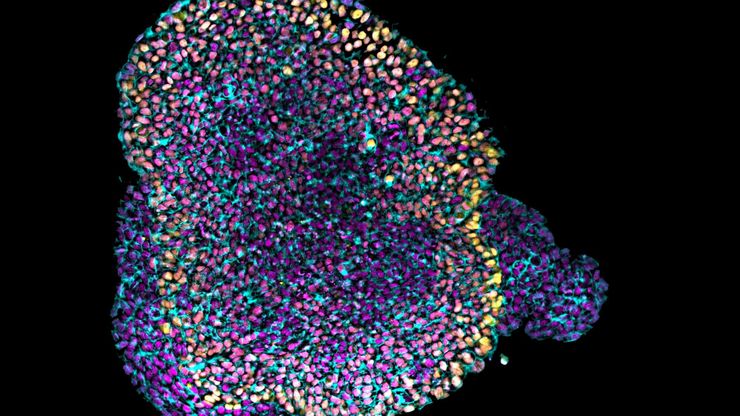
Volumetric Imaging of Cleared Organoids with Light Sheet
Cleared organoid imaging with Viventis Deep light sheet: rapid, high-resolution imaging of large organoids throughout their full volume.
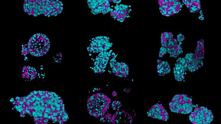
What’s the Best Organoid Imaging Approach for Early Drug Discovery?
Organoids and other complex in vitro models (CIVMs) are becoming increasingly important in early drug discovery and translational research, driven by the need for more predictive, human-relevant data…
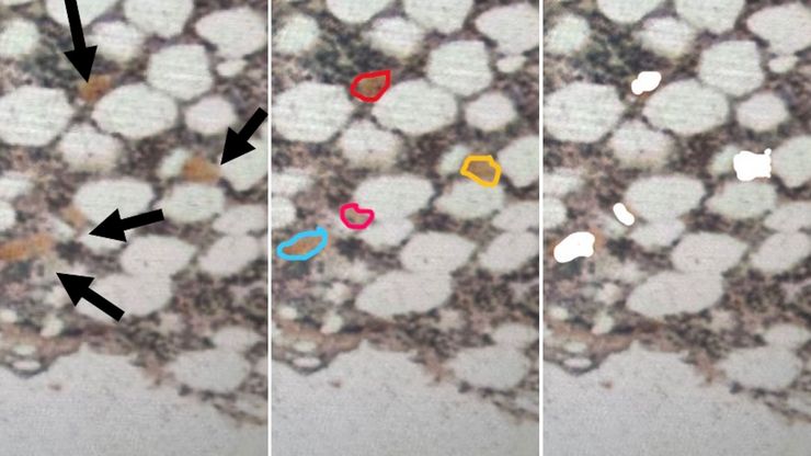
Spatial Proteomics Workflow in Blood Cancer (MPNs)
Megakaryocytes play a central role in the biology of myeloproliferative neoplasms (MPNs), yet their in vivo proteomic characterization remains a major challenge due to low abundance and disrupted…
History, Developments and Trends of Microscopy in Cancer Research
Cancer is a global disease, with 18 million new cases diagnosed and 10 million cancer-related deaths worldwide in 2020. This burden is set to increase, with a projected increase in cases of ~55% by…
Researchers Insights: Microscopy in Cancer Research
Discover how imaging techniques are driving cancer research forward. In this issue, we present comprehensive multimodal studies using microscopy, as well as new directions in intraoperative cancer…
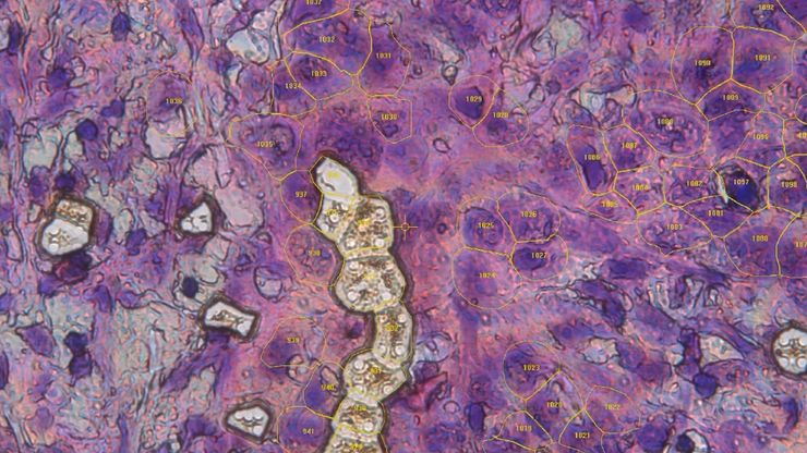
AI meets Deep Visual Proteomics (DVP) to Advance Disease Research
In this webinar, Dr. Andreas Mund will introduce a cutting-edge platform that merges Deep Visual Proteomics (DVP) with AI-powered pathology models, enabling high-resolution mapping of key regions in…
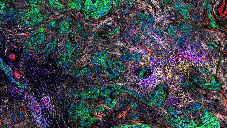
AI-Powered Hi-Plex Spatial Analysis Tools for Breast Cancer Research
Breast cancer (BC) is the leading cause of cancer-related deaths in women. Investigating the tumor microenvironment (TME) is crucial to elucidate the mechanisms of tumor progression. Systematic…
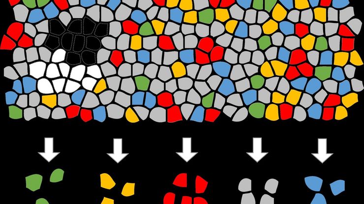
Biomarker Discovery with Laser Microdissection
Explore the potential of spatial proteomics workflows, such as Deep Visual Proteomics (DVP), to decipher pathology mechanisms and uncover druggable targets.
Altered protein expression, abundance, or…


