
Sciences de la vie
Sciences de la vie
C'est ici que vous pourrez développer vos connaissances, vos capacités de recherche et les applications pratiques de la microscopie dans divers domaines scientifiques. Apprenez à obtenir une visualisation précise, à interpréter les images et à faire progresser la recherche. Trouvez des informations pertinentes sur la microscopie avancée, les techniques d'imagerie, la préparation des échantillons et l'analyse des images. Les sujets abordés comprennent la biologie cellulaire, les neurosciences et la recherche sur le cancer, en mettant l'accent sur les applications et les innovations de pointe.
 tissue.")
AI-Powered Multiplexed Image Analysis to Explore Colon Adenocarcinoma
In this application note, we demonstrate a spatial biology workflow via an AI-powered multiplexed image analysis-based exploration of the tumor immune microenvironment in colon adenocarcinoma.

Laser Microdissection Protocols for Tissue and Cell Isolation - Download free eBook
In this Bio-protocol Selections, we present a collection of open-access, detailed methods papers using LCM to purify and isolate tissues and cells from plants, mouse embryos, cancer cells, neurons,…

How do Cells Talk to Each Other During Neurodevelopment?
Professor Silvia Capello presents her group’s research on cellular crosstalk in neurodevelopmental disorders, using models such as cerebral organoids and assembloids.
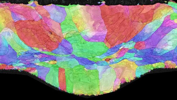
Workflow Solutions for Sample Preparation Methods for Material Science
This brochure presents and explains appropriate workflow solutions for the most frequently required sample preparation methods for material science samples.
.")
Dual-View LightSheet Microscope for Large Multicellular Systems
Visualizing the dynamics of complex multicellular systems is a fundamental goal in biology. To address the challenges of live imaging over large spatiotemporal scales, Franziska Moos et. al. present…

A Meta-cancer Analysis of the Tumor Spatial Microenvironment
Learn how clustering analysis of Cell DIVE datasets in Aivia can be used to understand tissue-specific and pan-cancer mechanisms of cancer progression
 tissue.")
Mapping the Landscape of Colorectal Adenocarcinoma with Imaging and AI
Discover deep insights in colon adenocarcinoma and other immuno-oncology realms through the potent combination of multiplexed imaging of Cell DIVE and Aivia AI-based image analysis

IBEX, Cell DIVE, and RNA-Seq: A Multi-omics Approach to Follicular Lymphoma
In a recent study by Radtke et al., a multi-omics spatial biology approach helps shed light on early relapsing lymphoma patients

Overcoming Observational Challenges in Organoid 3D Cell Culture
Learn how to overcome challenges in observing organoid growth. Read this article and discover new solutions for real-time monitoring which do not disturb the 3D structure of the organoids over time.
