
Sciences de la vie
Sciences de la vie
C'est ici que vous pourrez développer vos connaissances, vos capacités de recherche et les applications pratiques de la microscopie dans divers domaines scientifiques. Apprenez à obtenir une visualisation précise, à interpréter les images et à faire progresser la recherche. Trouvez des informations pertinentes sur la microscopie avancée, les techniques d'imagerie, la préparation des échantillons et l'analyse des images. Les sujets abordés comprennent la biologie cellulaire, les neurosciences et la recherche sur le cancer, en mettant l'accent sur les applications et les innovations de pointe.

Multiplexed Imaging Types, Benefits and Applications
Multiplexed imaging is an emerging and exciting way to extract information from human tissue samples by visualizing many more biomarkers than traditional microscopy. By observing many biomarkers…

How to Prepare your Specimen for Immunofluorescence Microscopy
Immunofluorescence (IF) specimen preparations can be analyzed by various fluorescence microscopy techniques, depending on the research application. IF is indispensable for researchers using a simple…

Designing your Research Study with Multiplexed IF Imaging
Multiplexed tissue analysis is a powerful technique that allows comparisons of cell-type locations and cell-type interactions within a single fixed tissue sample. It is common for researchers to ask…

Be Confident in your Results with Cell DIVE Validated Antibodies
The Cell DIVE System includes a carefully curated list of hundreds of commercially available antibodies validated to offer optimal specificity and sensitivity in multiplexed imaging. That validation…
 - THUNDER Imager 3D Cell Culture Influenca virus – red, cilia – green, Nuclei – blue.")
How Can Immunofluorescence Aid Virology Research?
Modern virology research has become as crucial now as ever before due to the global COVID-19 pandemic. There are many powerful technologies and assays that virologists can apply to their research into…

Chronic Inflammation Under the Microscope
In the course of chronic inflammation certain body areas are recurrently inflamed. This goes along with many human diseases. With the help of widefield light microscopy, the underlying processes can…
BABB Clearing and Imaging for High Resolution Confocal Microscopy
Multipohoton microscopy experiment using Leica TCS SP8 MP and Leica 20x/0.95 NA BABB immersion objective.
Understanding kidney microanatomy is key to detecting and identifying early events in kidney…
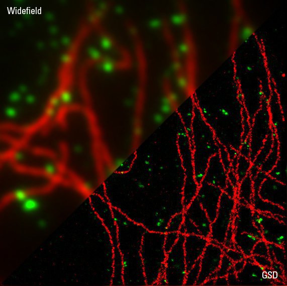
Sample Preparation for GSDIM Localization Microscopy – Protocols and Tips
The widefield super-resolution technique GSDIM (Ground State Depletion followed by individual molecule return) is a localization microscopy technique that is capable of resolving details as small as…
