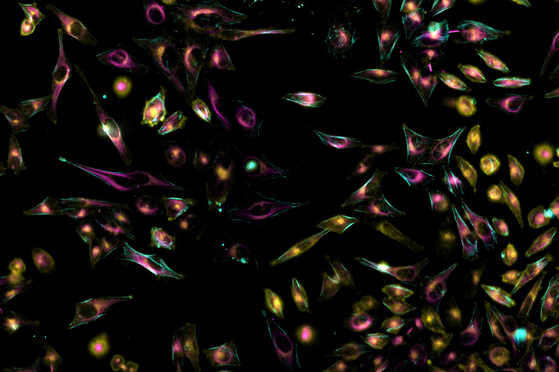
, SPY-Actin (cyan), and SiR-Tubulin (magenta). Instant Computational Clearing (ICC) was applied.")
Fast Live-Cell Imaging
Live-cell imaging is often utilized to discover intracellular processes. For example, transport processes, cytoskeleton dynamics, or mitosis, to name just a few. The according cellular structures are either tagged with fluorescent proteins or stained with live-cell dyes. Also, dynamic indicators like pH-sensors, Ca2+, or voltage sensitive dyes are available. Sometimes, also brightfield microscopy is an option.
Challenges
Fast intracellular events can be challenging for live-cell microscopy in terms of the following technical limitation. The temporal correlation of dynamic multicolor time-lapse imaging relies on the capability of the imaging system to record different fluorescent channels at the same time. Imagine a plane with one red and one green light flying in the sky. If your imaging system is only capable of recording one of the lights (red OR green) at a time, the plane will move forward in between the two consecutive images. Consequently, in a time-lapse movie you will not be able to tell if this is one (a red- and green-light plane), or two different planes (one red-light or one green-light plane).
Transferring this limitation to multicolor time-lapse imaging means, that you would not be able to properly correlate signals of two different fluorescence signals, e.g., a cargo protein and a motor protein of a fast-moving vesicle. This artifact is known as a spatiotemporal mismatch and is often an inherent problem of sequential imaging.
Classical fluorescence filter-based imaging systems suffer from the technical limitation described above, because changing filter cubes for the different fluorophores takes time.
Mica with its FluoSync™ technology (Figure 1) can acquire up to four fluorescence channels simultaneously. With it, 100% spatiotemporal correlation of four different cellular elements can be achieved. Additionally, exposure times of single channels do not add up with a simultaneous approach. Thus, the overall time consumption to acquire one time-point of a time-lapse experiment is shortened compared to sequential acquisition. Furthermore, Mica is an incubator which keeps cells at a given temperature and CO2 level over long time periods to have near-physiological imaging conditions.

Methods
Living cells were cultured either in Petri dishes or 96-well plates. For live-cell imaging, Mica was operated at 37°C, 5% CO2, and high humidity (to avoid concentration increases of solvents due to water evaporation). Imaging parameters are mentioned in the paragraphs below.
Results
In the following paragraphs, there are a few examples of live-cell imaging with Mica. Starting with an example showing the advantage of simultaneous imaging, over widefield THUNDER acquisition, through to high-speed confocal imaging.
1. Simultaneous vs. Sequential Imaging in Confocal mode
Videos 1a) and 1b) show the difference between sequential and simultaneous imaging. The same vesicle population was stained with two different live-cell dyes (green and red), meaning that all vesicles should appear yellow – the overlay of green and red.
Video 1a) shows both channels taken sequentially, one after the other. In this case, several vesicles appear to be either red or green, although they are both. It can be extremely difficult to differentiate red or green stationary vesicles from double stained moving ones.
In the Video 1b), both channels were acquired simultaneously. Here, all vesicles appear yellow due to the overlay of red and green.
2. Two-Color Time-Lapse Imaging – Widefield vs. THUNDER
In Video 2, HeLa cells were stained with WGA-Alexa Fluor™ 488 and SiR-Tubulin. The single images of the 3D time-lapse were taken every 5 seconds in widefield mode for a total duration of 2 minutes. Total z-stack size was 1.1 µm consisting of 6 layers.
The appropriate THUNDER method can be started and then synchronized with image acquisition or executed post acquisition.
3. Three-Color Time-Lapse Imaging – Widefield vs. THUNDER
In addition to WGA-488 and SiR-Tubulin, HeLa cells were now labelled with SPY-Actin. In this case, the 3D time-lapse imaging was recorded for 2 minutes with time intervals of 9 seconds and a total z-stack size of 1.8 µm divided into 9 layers.
4. Four-Color Time-Lapse Imaging – Confocal
Simultaneous imaging is also available for Mica confocal mode. In the case of Video 4, U2OS cells were labelled with MitoTracker™ Blue (blue), FluoView 488 Tubulin (green), WGA-Alexa555 (yellow), and SiR-Actin (cyan).
5. Four-Color Time-Lapse Imaging – THUNDER – High Speed
For Video 5, U343 cells were labelled with MitoTracker™ Deep Red and NucBlue™ in addition to the intrinsic marker tfLC3 (Tandem fluorescent-tagged LC3). TfLC3 is a fusion protein containing mRFP and eGFP, both of which show stability at different pH values. This leads to a redshift in more acidic environments, because mRFP is more stable at lower pH than eGFP. At neutral and higher pH, both fluorescent proteins are intact and thus, the resulting signal is yellow.
Conclusions
For 100% spatiotemporal correlation, fast cellular events demand simultaneous imaging of all channels which shall be acquired. Otherwise, signals from the channels are acquired at different time points and might lead to misinterpretations of dynamic multicolor time-lapse images.
Mica enables users to acquire the signal of up to four fluorophores at a time, either in confocal or widefield mode, including LIGHTNING and THUNDER. With it, cellular components can be imaged at the very same point in time, without spatiotemporal mismatch.
 and phalloidin (magenta), imaged using Viventis SCAPE; scale bar 50μm. Courtesy of Marina Cuenca and Heleen Jungen (Dayton lab), EMBL Barcelona.")
 at 2 weeks. Image acquired using Mica.")

