Christoph Greb , Dr.

Christoph Greb studied cell biology, parasitology and virology at the Philipps University in Marburg. In the course of his diploma thesis and his dissertation at the local Institute for Cytobiology and Cytopathology he examined the vesicular transport of apically destined proteins in polarized epithelial cells utilizing biochemistry as well as TIRF and confocal microscopy. From December 2011 he was writing for the Leica Science Lab as a freelancer. After his engagement for Novartis Vaccines & Diagnostics he started as Scientific Writer for the widefield team of Leica Microsystems in October 2013.
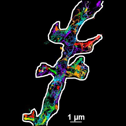
, tubulin with Cy5 (red), and nuclei with DAPI (blue). Image courtesy of Dr. Günter Giese, Max Planck Institute for Medical Research, Heidelberg, Germany.")
Overview of Fluorescent Dyes in terms of Applications and Properties
An introduction to commonly used fluorescent dyes and an overview of their characteristics are given in this article. Fluorescence microscopy is used for the study of specific cellular components with…

Infinity Optical Systems - From “Infinity Optics” to the Infinity Port
“Infinity Optics” is the concept of a light path with parallel rays between the objective and tube lens of a microscope [1]. Placing flat optical components into this “infinity space” which do not…
 segmentation used in conjunction with LMD to increase discovery throughput.")
Biomarker Discovery with Laser Microdissection
Explore the potential of spatial proteomics workflows, such as Deep Visual Proteomics (DVP), to decipher pathology mechanisms and uncover druggable targets.
Altered protein expression, abundance, or…

Deep Visual Proteomics Provides Precise Spatial Proteomic Information
Despite the availability of imaging methods and mass spectroscopy for spatial proteomics, a key challenge that remains is correlating images with single-cell resolution to protein-abundance…
 microdissected with a 10x objective (upper right). Inspection of the collection device (lower right).")
Molecular Biology Analysis facilitated with Laser Microdissection (LMD)
Extracting biomolecules, proteins, nucleic acids, lipids, and chromosomes, as well as extracting and manipulating cells and tissues with laser microdissection (LMD) enables insights to be gained into…

Technical Terms for Digital Microscope Cameras and Image Analysis
Learn more about the basic principles behind digital microscope camera technologies, how digital cameras work, and take advantage of a reference list of technical terms from this article.

Studying Virus Replication with Fluorescence Microscopy
The results from research on SARS-CoV-2 virus replication kinetics, adaption capabilities, and cytopathology in Vero E6 cells, done with the help of fluorescence microscopy, are described in this…
")
Introduction to Fluorescent Proteins
Overview of fluorescent proteins (FPs) from, red (RFP) to green (GFP) and blue (BFP), with a table showing their relevant spectral characteristics.

What is the Patch-Clamp Technique?
This article gives an introduction to the patch-clamp technique and how it is used to study the physiology of ion channels for neuroscience and other life-science fields.

Differential Interference Contrast (DIC) Microscopy
This article demonstrates how differential interference contrast (DIC) can be actually better than brightfield illumination when using microscopy to image unstained biological specimens.

Going Beyond Deconvolution
Widefield fluorescence microscopy is often used to visualize structures in life science specimens and obtain useful information. With the use of fluorescent proteins or dyes, discrete specimen…
 cells taken with phase contrast.")
Phase Contrast and Microscopy
This article explains phase contrast, an optical microscopy technique, which reveals fine details of unstained, transparent specimens that are difficult to see with common brightfield illumination.
Immersion Objectives
How an immersion objective, which has a liquid medium between it and the specimen being observed, helps increase the numerical aperture and microscope resolution is explained in this article.
 of an image of two points where the distance between them corresponds to the Rayleigh criterion.")
Microscope Resolution: Concepts, Factors and Calculation
This article explains in simple terms microscope resolution concepts, like the Airy disc, Abbe diffraction limit, Rayleigh criterion, and full width half max (FWHM). It also discusses the history.
, SPY-Actin (cyan), and SiR-Tubulin (magenta). Instant Computational Clearing (ICC) was applied.")
How to Perform Dynamic Multicolor Time-Lapse Imaging
Live-cell imaging sheds light on diverse cellular events. As many of these events have fast dynamics, the microscope imaging system must be fast enough to record every detail. One major advantage of…

RNA Quality after Different Tissue Sample Preparation
The influence of sample preparation and ultraviolet (UV) laser microdissection (UV LMD) on the quality of RNA from murine-brain tissue cryo-sections is described in this article. To obtain good…

3D Tissue Imaging: From Fast Overview To High Resolution With One Click
3D Tissue imaging is a widespread discipline in the life sciences. Researchers use it to reveal detailed information of tissue composition and integrity, to make conclusions from experimental…

How To Perform Fast & Stable Multicolor Live-Cell Imaging
With the help of live-cell imaging researchers gain insights into dynamic processes of living cells up to whole organisms. This includes intracellular as well as intercellular activities. Protein or…

Microscopy in Virology
The coronavirus SARS-CoV-2, causing the Covid-19 disease effects our world in all aspects. Research to find immunization and treatment methods, in other words to fight this virus, gained highest…

How to Sanitize a Microscope
Due to the current coronavirus pandemic, there are a lot of questions regarding decontamination methods of microscopes for safe usage. This informative article summarizes general decontamination…

Introduction to Mammalian Cell Culture
Mammalian cell culture is one of the basic pillars of life sciences. Without the ability to grow cells in the lab, the fast progress in disciplines like cell biology, immunology, or cancer research…

Introduction to Widefield Microscopy
This article gives an introduction to widefield microscopy, one of the most basic and commonly used microscopy techniques. It also shows the basic differences between widefield and confocal…

Photoactivatable, Photoconvertible, and Photoswitchable Fluorescent Proteins
Fluorescent proteins (FPs) such as GFP, YFP or DsRed are powerful tools to visualize cellular components in living cells. Nevertheless, there are circumstances when classical FPs reach their limits.…
 illuminated with wide-band UV excitation. Note the tissue structure with blue autofluorescence. Right: Same tissue and same immunostaining with FITC label illuminated with epi-illumination using narrow")
Milestones in Incident Light Fluorescence Microscopy
Since the middle of the last century, fluorescence microscopy developed into a bio scientific tool with one of the biggest impacts on our understanding of life. Watching cells and proteins with the…

Chronic Inflammation Under the Microscope
In the course of chronic inflammation certain body areas are recurrently inflamed. This goes along with many human diseases. With the help of widefield light microscopy, the underlying processes can…

Gene Editing with CRISPR/Cas9 - Breakthrough in Genome Engineering
The CRISPR/Cas9 system is one of several different bacterial systems for defense against viral attacks. It consists of two main components. One is a small piece of RNA which binds to the viral target…
Universal PAINT – Dynamic Super-Resolution Microscopy
Super-resolution microscopy techniques have revolutionized biology for the last ten years. With their help cellular components can now be visualized at the size of a protein. Nevertheless, imaging…
Image Processing for Widefield Microscopy
Fluorescence microscopy is a modern and steadily evolving tool to bring light to current cell biological questions. With the help of fluorescent proteins or dyes it is possible to make discrete…
Basic Principles of Luminescence
There are a lot of light-emitting processes occurring in nature. Luminescence is an umbrella term for those kinds of events where light emission is not the result of high temperatures. This article…

Fluorescent Proteins - From the Beginnings to the Nobel Prize
Fluorescent proteins are the fundament of recent fluorescence microscopy and its modern applications. Their discovery and consequent development was one of the most exciting innovations for life…
