")
Technical innovations in SpectraPlex
- Advanced spectral unmixing:
SpectraPlex employs cutting-edge unmixing algorithms that enable precise separation of overlapping fluorescent signals in multiplex experiments. This allows researchers to detect and visualize 8-15+ markers simultaneously within a single biological sample, offering greater insights without increasing complexity. - Enhanced STELLARIS confocal platform:
- White Light Laser (WLL): The WLL, spanning 440-790 nm, provides flexible excitation of multiple fluorophores, alongside a 405 nm laser for additional versatility. This flexibility expands the palette of usable fluorescent dyes and optimizes spectral separation.
- Photon-Counting Detectors: The inclusion of up to five detectors enables the detection of emissions between 410-850 nm, ensuring high sensitivity and a broad detection range.
- High-Resolution 3D Optical Sectioning: SpectraPlex enhances the platform’s ability to perform optical sectioning, delivering sharp, high-resolution 3D imaging, critical for studying intricate tissue structures or cellular interactions.
- Streamlined workflow:
The simplified workflow, featuring tools like the "Virtual Fridge" for fluorophore selection and automated reference data collection, drastically reduces setup time and minimizes manual errors during experiments. - Flexible approaches to spectral unmixing. SpectraPlex supports multiple strategies for data acquisition:
- Single stains for maximal precision.
- Group stains to conserve samples and resources.
- Theoretical spectra for semi-blind unmixing, enhancing flexibility for diverse applications.
")
Integration with Aivia software:
The pairing of SpectraPlex with Aivia delivers a robust solution for high-plex spatial phenotyping, enabling efficient and accurate workflows from imaging to data analysis.
Seamless data compatibility:
Imaging data generated by SpectraPlex is fully compatible with Aivia, Leica Microsystems’ advanced analysis software. This ensures researchers can immediately process their unmixed data for downstream analyses without requiring additional data formatting.
Advanced analytical tools:
Aivia provides powerful tools for spatial and quantitative analysis of multiplex imaging data. Researchers can map cellular phenotypes, examine spatial relationships, and explore disease mechanisms with greater depth and precision.
Automation & error reduction:
The integration includes intelligent automation to minimize human error and streamline data management. This combination helps researchers focus on their scientific insights rather than troubleshooting technical issues.
Applications:
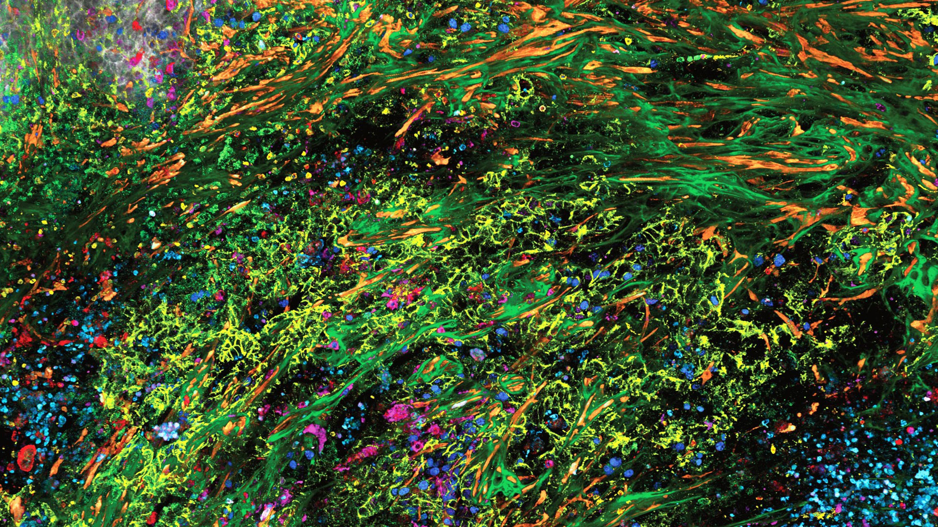
SpectraPlex and Aivia together have been demonstrated in diverse biological contexts, such as identifying cell types and functional markers in tissue (e.g., adenocarcinoma and cervical cancer models), revealing both spatial and temporal dynamics in 3D imaging.

.")

