
Multicolor Imaging with STELLARIS Confocal Platform
The STELLARIS family of confocal microscopes is equipped with next generation white light lasers (WLL). Paired with Power HyD detectors, STELLARIS provides complete spectral freedom. You can perform multicolor experiments to fit your research needs without being limited by the instrument. STELLARIS can also add an additional dimension to multicolor imaging by using the lifetime-based properties of fluorescent dyes/proteins with the TauSense set of tools.
Cytoskeleton and membranes in live cell imaged with TauSTED
Image acquired with STELLARIS STED.


TauSTED 775 resolves the intricate cytoskeleton network labeled with SiR-tubulin (glow - Spyrochrome), and trafficking vesicles labeled with CF594 (cyan - Biotium).
Endocytic pathway and mitochondria dynamics
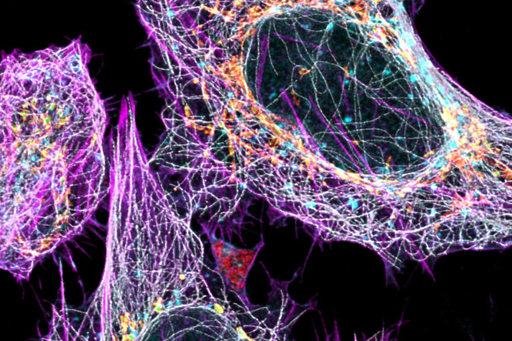
Cytoskeleton and membranes in live cell imaged with TauSTED
Image acquired with STELLARIS STED. Live cell TauSTED on U2OS cells, using labels for actin (SiR-actin, glow), microtubules (SPY555-tubulin, cyan), and membranes (CF488A coupled to WGA, green). SiR and SPY are available from Spirochrome. CF dyes are available from Biotium Inc. Scale bar: 10 μm


Cytoskeleton and membranes in live cell imaged with TauSTED
Root-hypocotyl junction of Arabidopsis thaliana

Cytoskeletal and mitochondrial interactions revealed
, AF750-Tom20 (760 – 790nm), AF790-Tubulin (810 – 850nm).")
Right: seen by STELLARIS 8 equipped with a Power HyD R detector. Spectral traces (right) show excitation (gray) and emission (red) spectra of fluorophores used in the experiment.
Sample courtesy: Dr.Jana Döhner and Dr.sc.nat. Urs Ziegler, University of Zürich.
Getting the most from live-cell experiments: fewer exposures, more information
When using traditional confocal microscopy to image several different fluorescent labels in the same sample, sequential imaging of each color channel is often needed to avoid spectral bleed-through, which can degrade image quality. In the case of a kinetic experiment, that means you may miss rapid dynamic events due to the increased time it takes to acquire each time point. In addition, your sample remains on the stage for longer, making it more challenging to maintain cell health for the duration the experiment. The image below was captured from live HeLa cells labeled with 4 different fluorophores to identify nuclei, actin, tubulin and plasma membrane. With STELLARIS, it was possible to collect all 4 channels in a single pass, rather than having to image the cells 4 times in succession.
, actin (yellow, SPY555), tublin (magenta, SPY650) and plasma membrane (gray, NIR750).")
to identify nuclei (cyan, Hoechst), actin (yellow, SPY555), tublin (magenta, SPY650) and plasma membrane (gray, NIR750).
LAS X Navigator – a GPS for your experiment

(shown on page X) labelled with Heidenhain azan trichrome stain. Transmitted
light detector (TLD) imaging was used to create an RGB overlay through excitation
with 488nm, 561nm and 638nm wavelengths.
Bottom: TauContrast image of the same field of view as above, maximum projection.
Discover at the speed of LIGHTNING
Intracellular components imaged with LIGHTNING

U2OS cells labeled with AF488 (microtubules, gray), SPY555 (actin, magenta), MitoTrackerRed (lumen of mitochondria, green), Atto 647N (TOM 20, Mitochondria, red), and CF770 WGA (membranes, cyan).


