
Why is multiplexing necessary?
Simple multicolor experiments utilize up to three different fluorophores with distinctive emissions in the blue, green, and red regions of the visible spectra. However, the increasing complexity of biological samples and processes has raised the technical and application-driven demand for methods that allow for simultaneous imaging in the mid-plex (>10 fluorophores) range [5-7].
Challenges
One way to achieve multiplexing is to perform iterative staining and imaging rounds on the sample using the above mentioned blue, green, and red fluorophores. However, this procedure is very time-consuming and can compromise sample integrity and quality due to the harsh treatments that are required during each round of staining. In addition, image processing to combine the data is cumbersome and particularly difficult in 3D tissue samples. Therefore, strategies to achieve the required multiplexing level in a single round of staining have become desirable for more complex multiplexing applications. While increasing the number of different fluorophores in a sample is a task that researchers can tackle, purely from a sample preparation point of view, dealing with the increased overlap of the excitation or emission spectra of multiple fluorophores poses a challenge even for experienced researchers. Cross excitation of fluorophores and crosstalk or bleed-through of the emission spectra make it particularly difficult to distinguish between the different signals. Each additional fluorophore on a sample therefore increases the challenge to properly separate each label.
Methods
The STELLARIS confocal platform was designed to serve these applications. The ultra-flexible spectral capabilities enabled by the next generation WLL super-continuum excitation, AOBS, optimized beam path , and up to 5 Power HyD detectors working simultaneously, ensure maximized photon collection coming from every relevant marker. Spectral unmixing tools integrated in the ImageCompass user interface make the separation of complex fluorophore combinations possible.
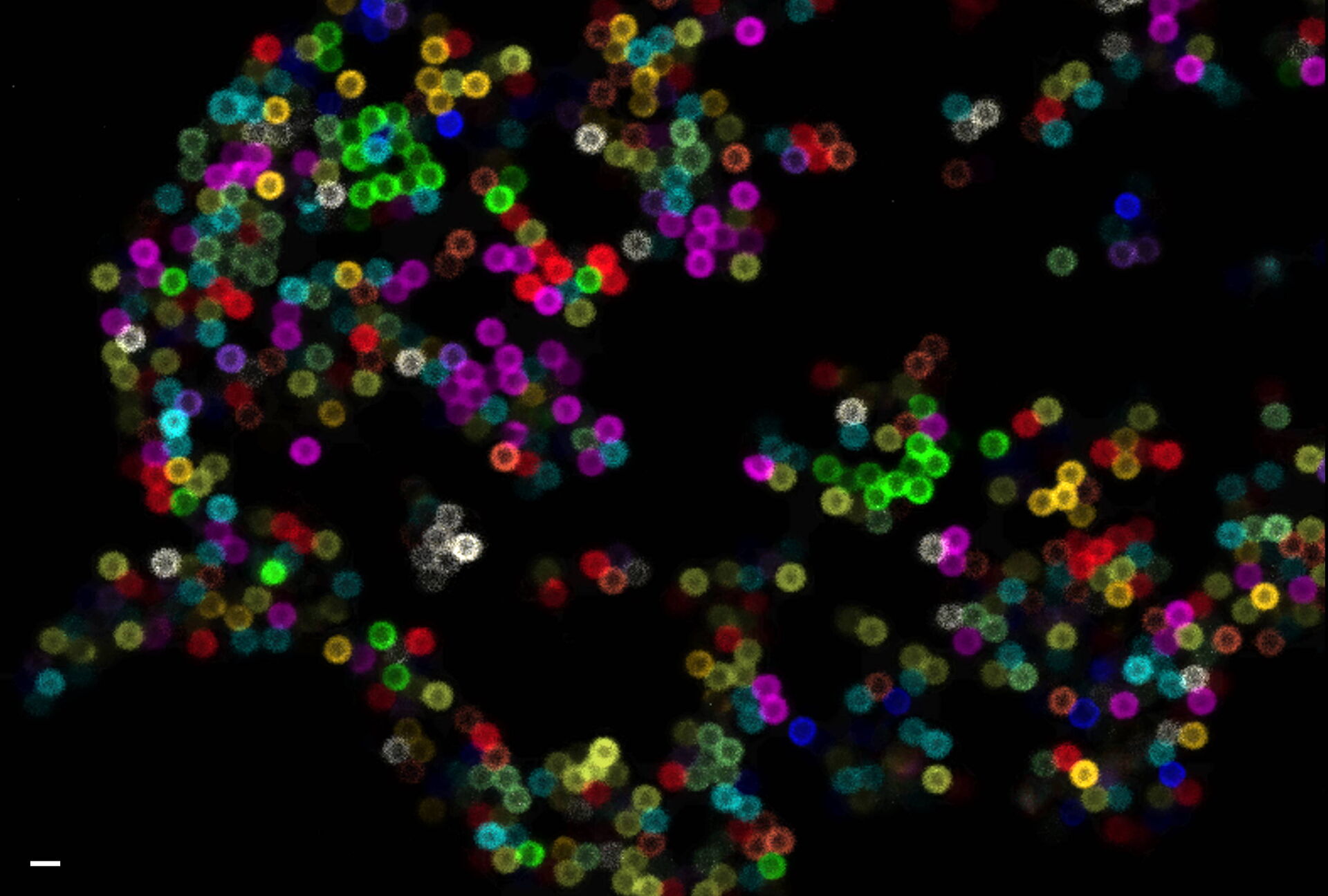
As an example, we outline one approach to image 11 different fluorophores in a single round of staining and imaging. We defined a set of spectrally different fluorophores and separated them by applying the linear unmixing, channel dye separation tool in STELLARIS. To illustrate how this works, we selected a panel of 11 Alexa Fluor (AF) dyes ranging from AF 405 nm to AF 790 nm (Figure 1), conjugated to streptavidin. We then coupled each fluorophore to biotin coated polystyrene beads, purified the beads, and mounted a mixture of them in Prolong Diamond on glass coverslips. The 11-color sample was imaged using a STELLARIS microscope equipped with 5 Power HyD detectors (Figure 2).
 and emission (B) spectra of Alexa Fluor dyes coupled to beads used in this article.")
Figure 1: Excitation (A) and emission (B) spectra of Alexa Fluor dyes coupled to beads used in this article. The spectra were generated with FPbase [8] (www.fpbase.org).
 Raw image of the 11-color beads (nominal diameter 0.8 micron) sample, and (B) image obtained with the dye separation tool integrated in STELLARIS.")
To assess the efficiency of the dye separation, we quantified the crosstalk of each fluorophore on the single beads within the different channels (Fig. 3). In the ideal case, each bead would appear in only one channel, as we labeled them with only one fluorophore. For quantification, we used the Maximum Entropy algorithm [9] embedded in the auto thresholding function of Fiji [10], segmented the objects (beads, Figure 3A) for each channel (shown as magenta in Figure 3B), and measured the normalized mean intensity in all channels. The normalized mean intensity values are displayed in the histograms (Figure 3C). Our analysis confirms that each fluorophore is assigned to one channel, and the 11 differently labeled beads can clearly be separated into their respective channels.

Results
The overall result was a successful spectral separation of 11 different fluorophores that were coupled to synthetic beads, exemplifying how the STELLARIS confocal platform provides a unique and powerful opportunity for multiplexing applications.
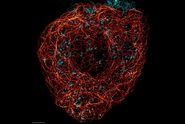

, actin network (ATTO 647N), and nuclear pore basket (CF 680R).")
