Related Articles

Overcoming Challenges with Microscopy when Imaging Moving Zebrafish Larvae
Zebrafish is a valuable model organism with many beneficial traits. However, imaging a full organism poses challenges as it is not stationary. Here, this case study shows how zebrafish larvae can be…
, SPY-Actin (cyan), and SiR-Tubulin (magenta). Instant Computational Clearing (ICC) was applied.")
How to Perform Dynamic Multicolor Time-Lapse Imaging
Live-cell imaging sheds light on diverse cellular events. As many of these events have fast dynamics, the microscope imaging system must be fast enough to record every detail. One major advantage of…

3D Tissue Imaging: From Fast Overview To High Resolution With One Click
3D Tissue imaging is a widespread discipline in the life sciences. Researchers use it to reveal detailed information of tissue composition and integrity, to make conclusions from experimental…

Simplifying Complex Fluorescence Multiwell Plate Assays
Apoptosis, or programmed cell death, occurs during organism embryo development to eliminate unwanted cells and during healing in adults to rid the body of damaged cells and help prevent cancer.…

Efficient Long-term Time-lapse Microscopy
When doing time-lapse microscopy experiments with spheroids, there are certain challenges which can arise. As the experiments can last for several days, prolonged sample survival must be achieved…

Using Machine Learning in Microscopy Image Analysis
Recent exciting advances in microscopy technologies have led to exponential growth in quality and quantity of image data captured in biomedical research. However, analyzing large and increasingly…
The AI-Powered Pixel Classifier
Achieving reproducible results manually requires expertise and is tedious work. But now there is a way to overcome these challenges by speeding up this analysis to extract the real value of the image…
, Astrocytes (GFAP, red), Nuclei (DAPI, blue).")
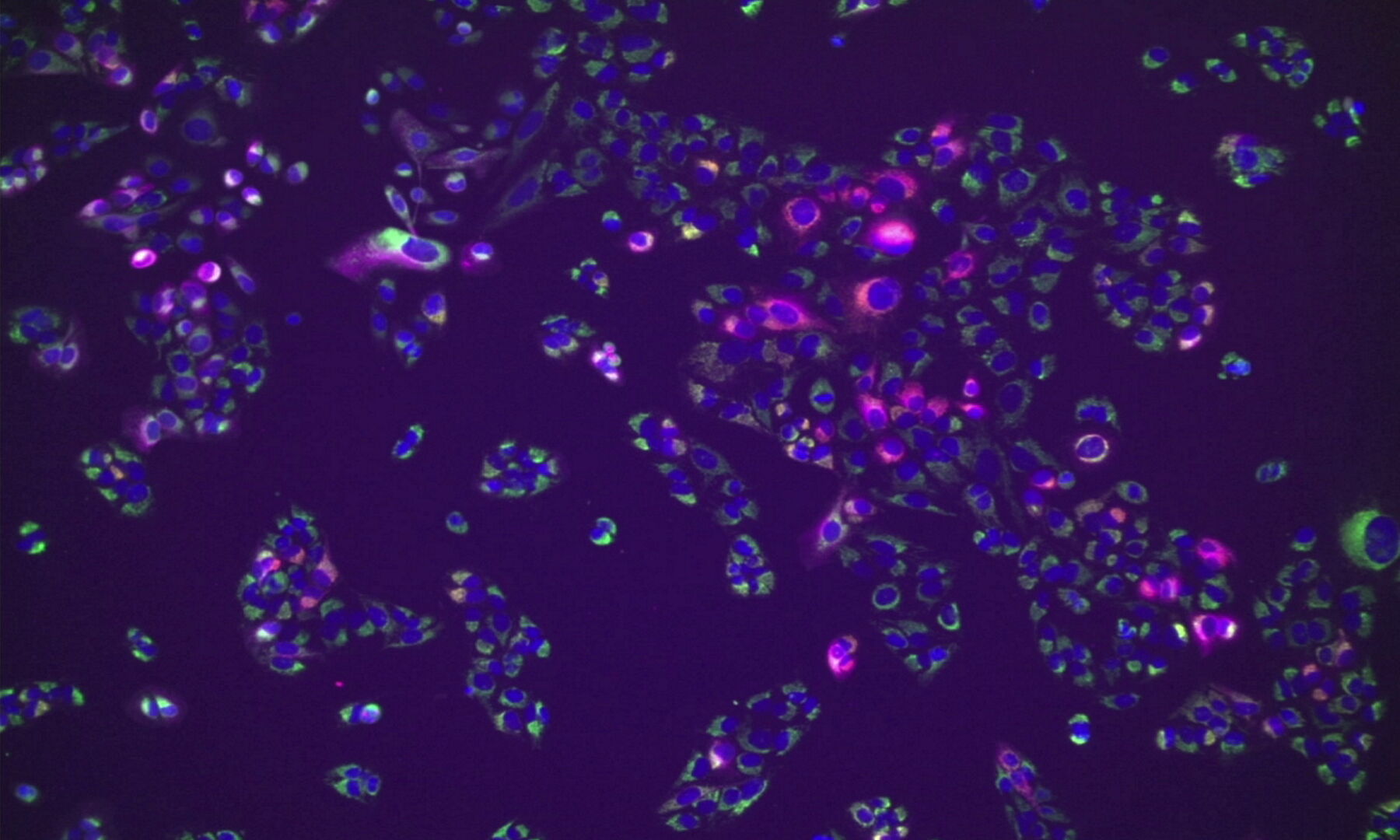
Multicolor Microscopy: The Importance of Multiplexing
The term multiplexing refers to the use of multiple fluorescent dyes to examine various elements within a sample. Multiplexing allows related components and processes to be observed in parallel,…
