
What you will learn
- Produce consistent, high‑quality ultrathin sections: The eBook explains how uniform section thickness underpins TEM, SEM, array tomography, and 3D reconstruction. It shows how section thickness correlates with interference colors and how balanced feed and cutting speed help generate stable, clean ribbons.
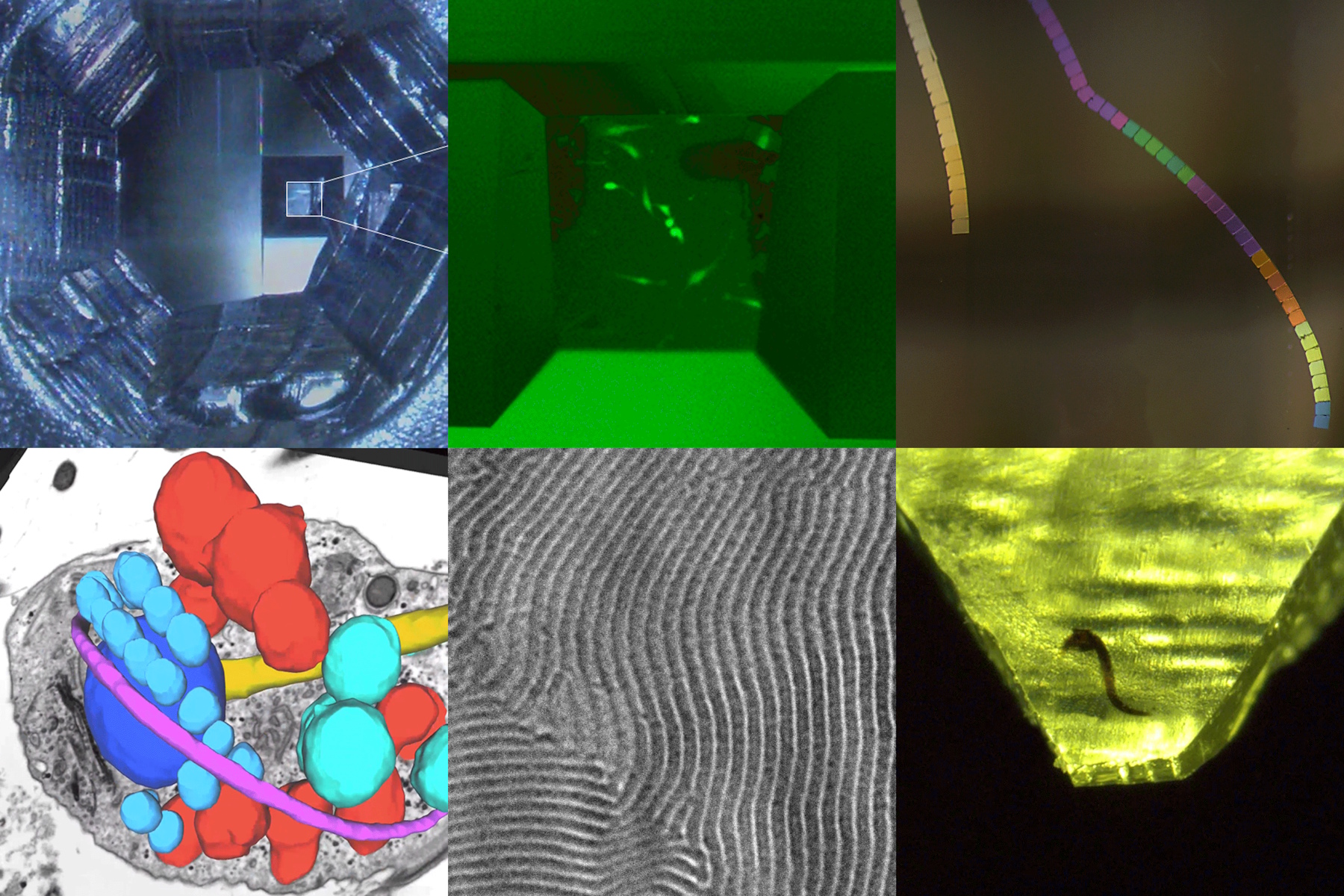
- Target labeled or hidden structures with confidence: Readers learn two modern strategies for reaching regions of interest: in‑resin fluorescence for visualizing labeled cells directly in the block face, and micro‑CT for mapping buried structures before trimming begins. These methods reduce guesswork and help expose targets efficiently.
- Streamline workflows through guided trimming and alignment: The eBook highlights how guided trimming routines and camera‑supported alignment help users prepare reproducible block faces and achieve reliable knife‑to‑sample geometry. This reduces variability and makes sectioning more accessible for new and occasional users.
- Apply ultramicrotomy across biology, materials science, and volume EM: From fluorescence‑guided CLEM workflows to polymer sectioning and serial sectioning for volume EM, the eBook provides practical examples that show how precise trimming, alignment, and sectioning extend across diverse samples and imaging goals.
- Build skills progressively with structured, real‑world guidance: Each chapter follows a clear sequence—context, application note, workflow takeaways—making it easy to adopt new techniques step by step, whether you're trimming toward a fluorescent target or preparing delicate polymers for cryo‑sectioning.
Why section quality matters in modern EM
Modern electron microscopy depends on producing consistent, high‑quality sections, yet ultramicrotomy has long been considered a specialist skill. As research increasingly relies on 3D datasets, CLEM workflows, and automated analysis pipelines, the need for reliable sectioning—across users and sample types—has never been greater. This eBook brings together practical application notes, expert insights, and real laboratory workflows to help users navigate today’s ultramicrotomy landscape.
Building the foundation: Understanding section thickness & ribbons
The early chapters provide a clear foundation in section quality. They describe how uniform thickness affects imaging fidelity and how interference colors offer a quick, intuitive readout of section thickness. Through examples and imagery, readers see how feed settings, sectioning speeds, and clean edge geometry contribute to producing stable ribbons suitable for high‑resolution EM and array tomography.
Smarter targeting: Fluorescence & micro‑CT workflows
The eBook then moves into strategies for targeted trimming. In‑resin fluorescence enables direct visualization of labeled cells or structures within the block face, eliminating the need for repeated semi‑thin sections. For structures buried deep within resin, 3D micro‑CT provides a volumetric map that guides trimming toward internal targets. Together, these approaches allow users to reach regions of interest more efficiently and with greater confidence, reducing wasted material and time.
Making workflows reproducible: Guided trimming & alignment
Beyond targeting, the eBook focuses on workflow reproducibility. Automated or guided trimming routines help users define block‑face dimensions and trimming depth, while camera‑supported alignment routines simplify one of the most delicate steps: bringing knife and block into perfect orientation. These workflow aids help both trainees and experienced microscopists achieve consistent results, making sectioning less dependent on individual technique.
Applications across EM, CLEM, cryo & polymers
A major strength of the resource is its diversity of applications. Readers gain insights into fluorescence‑guided CLEM, cryo workflows for preserving delicate structures, and the nuances of polymer sectioning—where material properties dictate whether room‑temperature or cryogenic methods are appropriate. Later chapters show how ultramicrotomy supports volume EM pipelines, where long, consistent ribbons enable large‑scale 3D reconstructions enhanced by AI‑based image analysis.
Practical guidance designed for real lab workflows
Throughout, the eBook’s structure—context, application note, key takeaways—makes it easy to translate knowledge into practice. Whether users are troubleshooting fundamental sectioning parameters, adopting fluorescence navigation, or preparing complex materials samples, the guidance empowers them to work more efficiently and confidently in the ultramicrotomy workflow.
