
How can AR improve visualization during microsurgery?
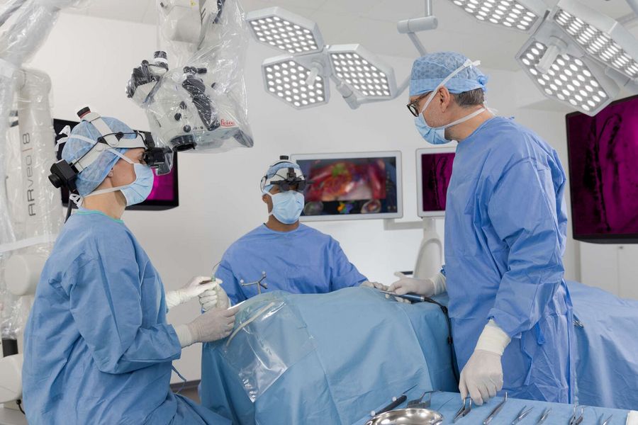
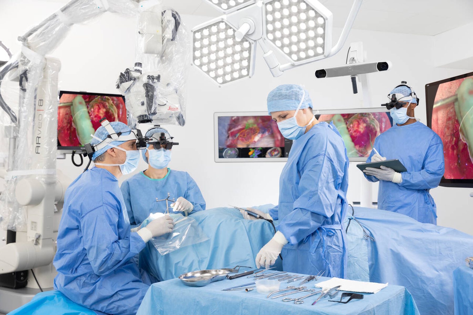
In the medical field, several tools are used for pre-operative investigations, including imaging techniques such as CT-scan, MRI, angiography, ultrasound, or transcranial magnetic stimulation. All this information can supplement a surgical procedure via 3D imaging technology. Surgeons can visualize the operating field with AR information on 3D visualization viewing systems such as 3D monitors or headsets. There is also a wide range of surgical visualization tools.
Operating microscopes, for example, provide magnification but also support fluorescence imaging (for example with 5-ALA), neuro-monitoring, neuro-mapping, neuro-navigation, and offer a Mixed Reality navigation viewing experience. The surgeon can access the operating field and AR overlays in a digitally enhanced live image, displayed on a monitor or a digital headset.
Related articles
Augmented Reality: Transforming Neurosurgical Procedures
 set-up.")
Digitalization in Neurosurgical Planning and Procedures

Benefits of Fluorescence in Vascular Neurosurgery

Augmented Reality (AR) Fluorescence Image Gallery
How AR Fluorescence Imaging Supports Neurovascular Surgery

Overcoming limitations in Vascular neurosurgery
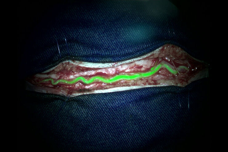
During the Leica Neurosurgical Visualization summit, Dr Raphael Guzman, Professor of Neurosurgery at the University of Basel in Switzerland, and Chair of the Department of Neurosurgery, spoke about fluorescence-guided neurosurgery and videoangiography.
Overcoming the limitations of Indocyanine Green (ICG), the GLOW800 AR fluorescence application from Leica Microsystems, provides augmented information in the surgical field, allowing anatomy assessment and perfusion of blood vessels. GLOW800 AR combines the white light image with real-time fluorescence imaging in a digitally processed image superimposed on the blood vessels.
Related articles
Aneurysm Clipping: Assessing Perforators in Real-Time with AR Fluorescence

How Augmented Reality is Transforming Vascular Neurosurgery
GLOW800 Augmented Reality Fluorescence in Aneurysm Treatment
How AR Helps in the Surgical Treatment of Moyamoya Disease

How AR supports Plastic Reconstructive Surgery
During oncological reconstructive surgery procedures, AR can help surgeons to accomplish the following:
- Confirm the viability of the flap with indocyanine green (ICG)
- Identify the viable mastectomy skin, as well as the nonviable skin and tissue.
- Increase the precision of the flap delimitation
- Avoid complications, such as fat necrosis
 and florescence-aided angiography FL800 (right). Image courtesy of Prof. Küntscher.")
Related articles
Using GLOW800 AR in Radial Forearm Flap Free Phalloplasty

How AR Surgery Benefits Radial Forearm Free Flap Phalloplasty

Free Flap Procedures in Oncological Reconstructive Surgery

Advances in Oncological Reconstructive Surgery

How AR supports uninterrupted workflow and helps improve ergonomics
During surgery, surgeons need to check multiple monitors to get information from different sources. With the use of AR, access to the information becomes easier, preventing the interruption of the surgical workflow.
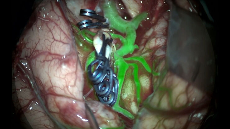
During aneurysm surgery for example, the GLOW800 AR fluorescence application allows surgeons to verify, in real-time, that all branches are open and perfused while the aneurysm is excluded from the circulation thanks to the GLOW AR view, showing green fluorescence in the blood vessels in a natural white light view of the cerebral anatomy.
By having that one single image, surgeons no longer need to look at different screens to get a read on the surgical area, allowing them to work uninterrupted.

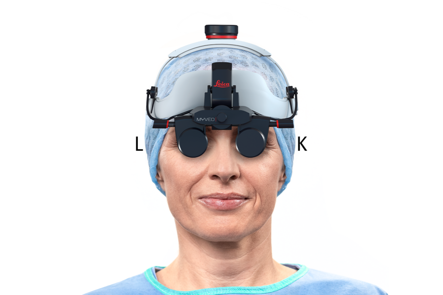
MyVeo all-in-one surgical visualization headset
Leica Microsystems has introduced the MyVeo all-in-one surgical visualization headset, freeing surgeons from the microscope by unifying essential clinical data right in front of their eyes in real time. Surgeons can now experience an entire surgery with the MyVeo headset, providing a single integrated view for an uninterrupted workflow, supporting more confident decision-making during surgery.
The MyVeo headset shows the normal white light image of the operating field as well as AR applications, image-guided surgery (IGS) data and even endoscope image feeds. Thanks to MyVeo, surgeons can now experience ergonomic comfort and ease of movement during long surgeries as they have the right view constantly in front of their eyes, achieving a comfortable upright working posture.
Choose from different viewing devices for 3D surgery
With the Evolved ARveo 8 digital visualization microscope, the OR team can choose from different viewing devices for 3D surgery and even use them interchangeably. 3D visualization can be achieved directly with the MyVeo headset, or via a 3D 55-inch 4K 3D cart-mounted external monitor for heads-up-display surgery. For a shared 3D view in the OR, the microscope-mounted 31-inch 4K 3D monitor shows the images to the entire OR staff.