3D high-plex imaging in Cancer Immunology. Detail of a pancreatic tumor section in mouse model, labeled with 15 markers and imaged in one go using STELLARIS SpectraPlex. Total imaged volume 3.2 mm x 2.2 mm x 70 mm in 1 h 20 min. Source: 3D high-multiplex imaging in cancer immunology. Kunz L., Speziale D., et al., Nat. Methods (2024).

Advanced Tissue Imaging & Analysis

To gain insight into biological processes and disease mechanisms, scientists examine tissues at multiple levels to understand how cells and extracellular components interact. Advanced imaging and analysis solutions help drive spatial biology research by providing detailed visualization and comprehensive analysis of tissues.

Simply get in touch!

Our imaging experts are here to help with advice on solutions for Advanced Tissue Imaging and Analysis.

Image acquired with TauSTED Xtend. 3 color STED sample. Cyan: Vimentin, AF594. Magenta: Phalloidin-Actin, Atto 647N. Glow: NUP 107,  CF680R. Sample Courtesy of: Dr. Mariano Gonzales Pisfil and Dr. Steffen Dietzel, Core Facility Bioimaging at the Biomedical Center Ludwig-Maximilians-Universität München, Germany.

Versatility for Every Application

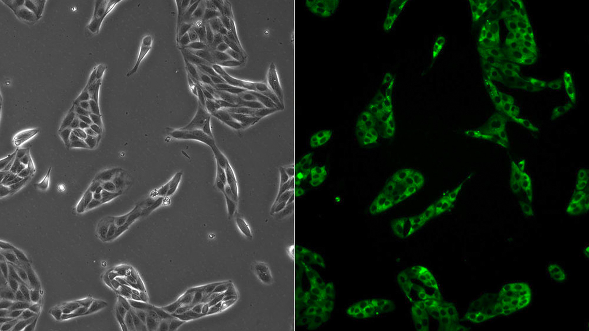
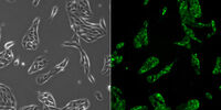
To acquire tissue data in both 2D and 3D, our imaging solutions range from widefield, for visualizing large tissue sections, to super-resolution confocal imaging, to reveal very fine tissue structures. This versatility ensures scientists have the right tools to study tissue structure and function effectively.

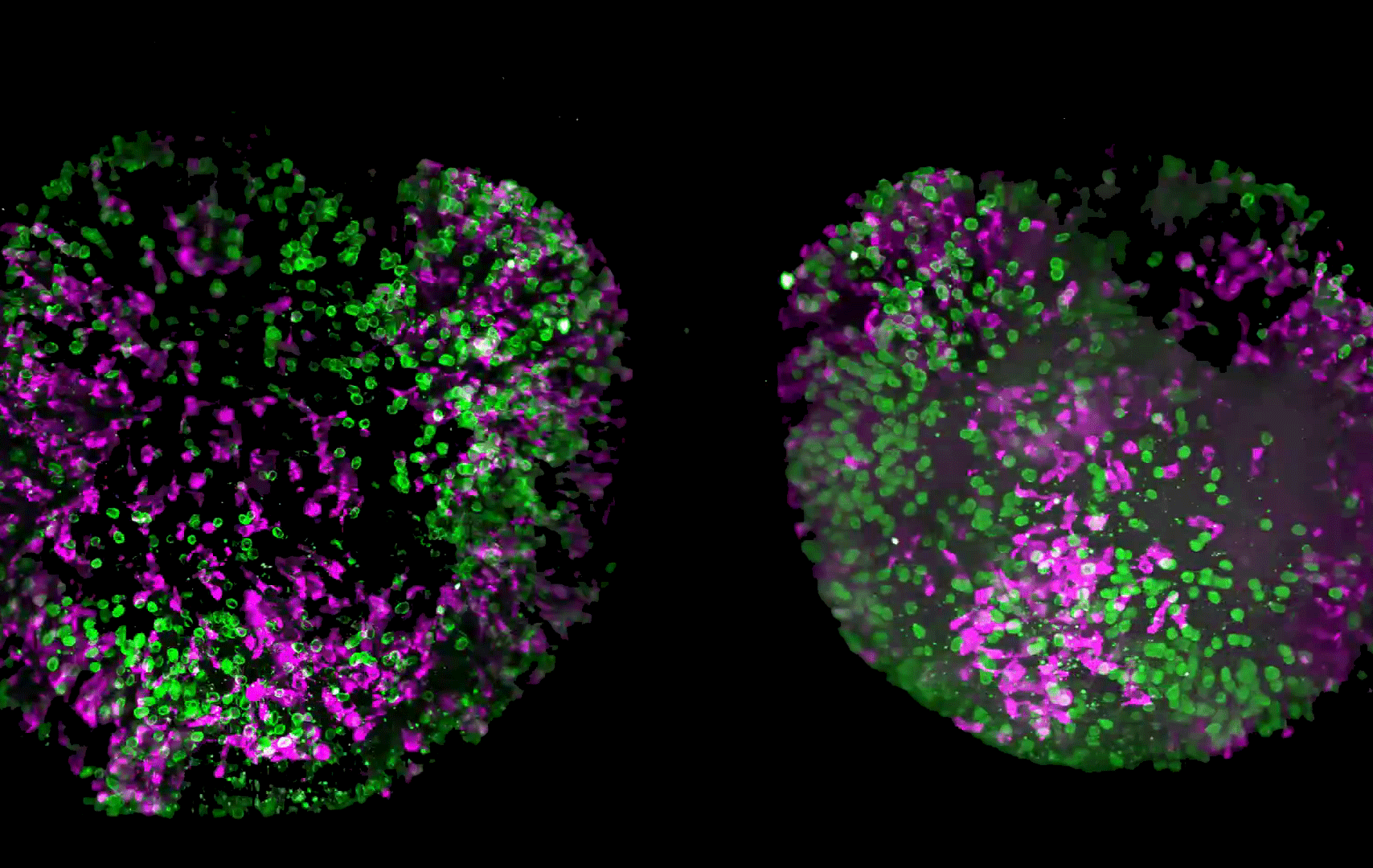
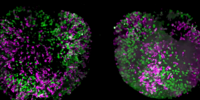
Advanced multiplexing and phenotyping

For spatial biology multiplexing and phenotyping, scientists can analyze multiple biomarkers within tissue samples using our multiplex solutions. They include automated iterative staining with 60+ biomarkers in one sample, which is essential for studying complex biological processes and interactions.

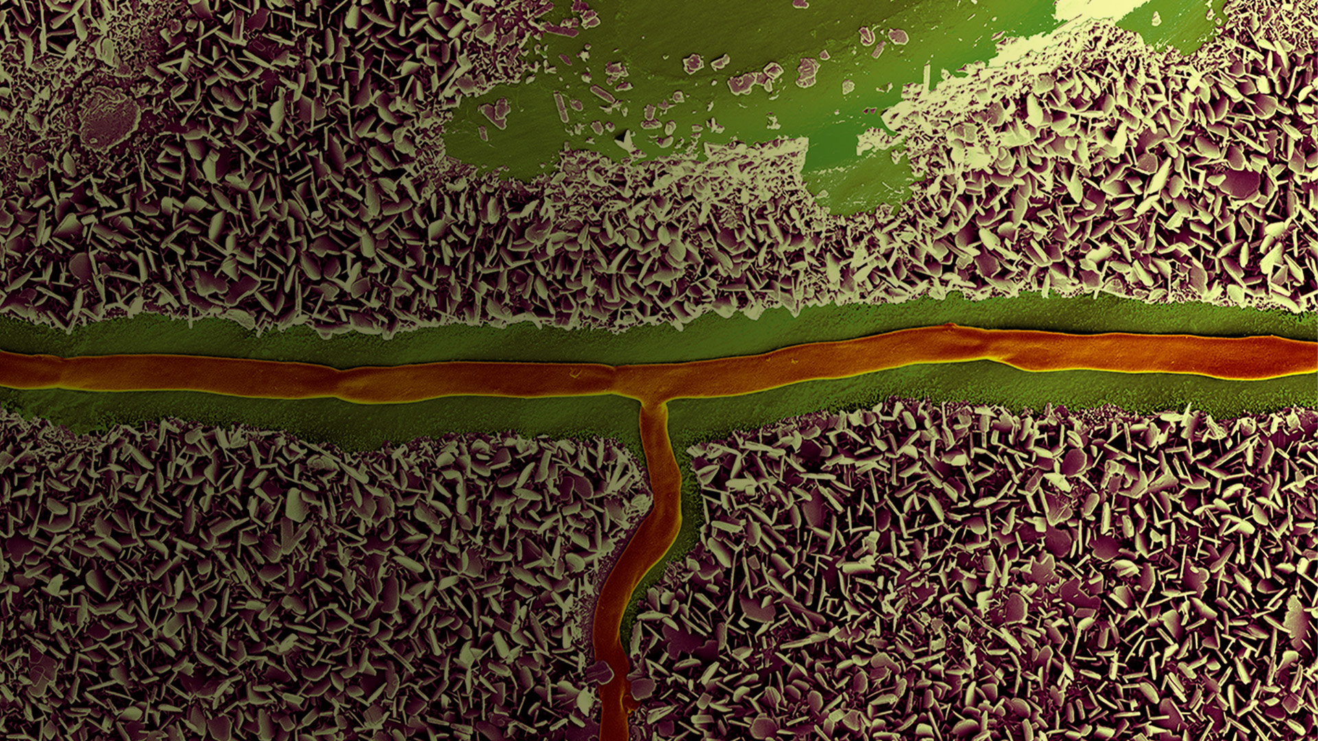
Comprehensive tissue scanning solutions

Take advantage of our multi-functional instruments, including those for thin section and volumetric scanning, to support a wide range of tissue analysis applications. This flexibility ensures the optimal solution can be applied to study tissue structure and function effectively.

What are the advantages of using Leica microscope solutions for tissue imaging & analysis?


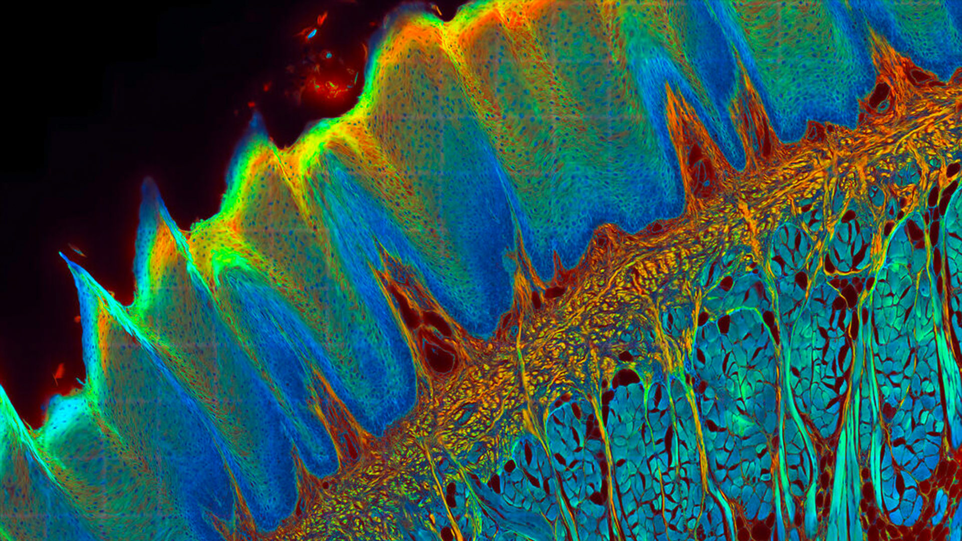
Why study tissue structure and function?

Scientists examine tissue structure and function across scales to better understand tissue, cellular, and molecular relationships. Leica imaging solutions and sample preparation techniques can be used to differentiate cells by their phenotypes and translate these insights into implications for health and disease.


What methods are best for studying tissue function?

It is essential to visualize and analyze biological processes within tissues for scientists to improve their understanding of how tissues function and respond to various stimuli. Leica functional imaging and spatial phenotyping tools make it easier for them to achieve this goal.


How can reproducible results be obtained rapidly?

Reliable, reproducible results come from standardized workflows and automated imaging combined with Aivia, our AI-powered analysis software, which accelerates data processing, reduces errors, and ensures consistent, high-quality insights.

Frequently asked questions about Advanced Tissue Imaging and Analysis

Show answer What is tissue structure and why is it important?

Tissue structure refers to the organization of cells and extracellular components within tissues. Understanding this architecture is essential for studying biological processes, disease mechanisms, and developing therapeutic strategies.

Show answer What are the main types of tissues in the human body?

There are four primary tissue types: 

  • Epithelial: Covers surfaces and lines cavities.
  • Connective: Provides structural support and transport.
  • Muscle: Enables movement through contraction.
  • Nervous: Facilitates communication via electrical impulses
Show answer How can tissue structure be analyzed in detail?

Researchers use advanced imaging techniques such as confocal microscopy, volumetric scanning, and ultramicrotomy to visualize tissue architecture at multiple scales, from whole tissue sections to subcellular resolution.

Show answer What methods are best for studying tissue function?

Functional imaging and spatial phenotyping allow scientists to examine biological processes within tissues, revealing how they interact at the molecular level.

Show answer How can reproducible results be achieved quickly?

Standardized workflows and AI analysis tools ensure consistency and speed, reducing variability and improving data reliability for tissue research.

Show answer What role does histology play in tissue research?

Histology, the study of tissues using staining and microscopy, is fundamental for identifying tissue morphology and diagnosing pathological changes. Techniques include chemical-based staining (e.g., H&E) and antibody-mediated staining for specific biomarkers.

Show answer Which imaging solutions support both 2D and 3D tissue analysis?

Leica Microsystems offers versatile platforms like STELLARIS confocal systems, Mica, Cell DIVE and SpectraPlex for high-resolution imaging in both 2D and 3D, enabling multiplex analysis and spatial biology workflows.

Show answer How does tissue research impact medical science?

Insights into tissue structure and function inform disease diagnosis, drug development, and regenerative medicine, making tissue biology a cornerstone of biomedical research.

Related Articles

Read our latest articles about Advanced Tissue Imaging and Analysis

The knowledge portal of Leica Microsystems offers scientific research and teaching material on the subjects of microscopy. The content is designed to support beginners, experienced practitioners and scientists alike in their everyday work and experiments.

More Articles
Final Segmentation of organelles in Trichomonas species. Magenta – costa, light blue – hydrogenosomes, turquoise – ER, red – vacuoles, yellow – axostyle, green – Golgi apparatus.  Sample courtesy of Isabelle Guerin-Bonne, Low Kay En, Electron Microscopy Unit, Yong Loo Lin School of Medicine, National University of Singapore. Scale bar: 1 µm.

Volume EM and AI Image Analysis

The article outlines a detailed workflow for studying biological tissues in three dimensions using volume-scanning electron microscopy (volume-SEM) combined with AI-assisted image analysis. The focus…
Pancreatic Ductal Adenocarcinoma imaged with Cell DIVE. Analysis done by Aivia.

A Guide to Spatial Biology

What is spatial biology, and how can researchers leverage its tools to meet the growing demands of biological questions in the post-omics era? This article provides a brief overview of spatial biology…
Masson-Goldner staining of a hedgehog brain slice.

How to Streamline Your Histology Workflows

Streamline your histology workflows. The unique Fluosync detection method embedded into Mica enables high-res RGB color imaging in one shot.
Spectral separation of 11 fluorophores coupled to polystyrene beads on a STELLARIS confocal system.

Multiplexing through Spectral Separation of 11 Colors

Fluorescence microscopy is a fundamental tool for life science research that has evolved and matured together with the development of multicolor labeling strategies in cells tissues and model…
3D Reconstruction of brain slide image_Mica

3D Tissue Imaging: From Fast Overview To High Resolution With One Click

3D Tissue imaging is a widespread discipline in the life sciences. Researchers use it to reveal detailed information of tissue composition and integrity, to make conclusions from experimental…

Tissue Image Gallery

Visual analysis of animal and human tissues is critical to understand complex diseases such as cancer or neurodegeneration. From basic immunohistochemistry to intravital imaging, confocal microscopy…
Brain organoids labeled with lamin (green) and tubulin (magenta), acquired using Viventis Deep Dual View Light Sheet Microscope. Courtesy of Akanksha Jain, Treutlein Lab ETH-DBSSE Basel (Switzerland).

Advanced Imaging Solutions for Biopharma

Advanced imaging solutions empower biopharma researchers to uncover critical insights in drug discovery, quality control, and understanding complex biological systems. For biopharma, Leica solutions help accelerate drug discovery, enhance cellular analysis, and support data integrity that meets regulations.

Read More

Cell Culture

Growing cells under lab conditions is the base for scientists working in fields of cell or developmental biology, cancer research, or any kind of life science and pharma research. Find out how Leica cell culture solutions PAULA, DMi1 and DM IL help to culture animal cells in the lab.

Read More

EM Sample Prep Workflows & Uses

Researchers can consistently achieve high-quality, precise, and reproducible results when imaging samples with electron microscopy by using Leica sample preparation solutions. Our solutions support Scanning Electron Microscopy (SEM), Transmission Electron Microscopy (TEM), and Cryo EM.

Read More

Organoids and 3D Cell Culture

One of the most exciting recent advancements in life science research is the development of 3D cell culture systems, such as organoids, spheroids, or organ-on-a-chip models. A 3D cell culture is an artificial environment in which cells are able to grow and interact with their surroundings in all 3 dimensions. These conditions are similar to how they would be in vivo.

Read More

Brain organoids labeled with lamin (green) and tubulin (magenta), acquired using Viventis Deep Dual View Light Sheet Microscope. Courtesy of Akanksha Jain, Treutlein Lab ETH-DBSSE Basel (Switzerland).
Scroll to top