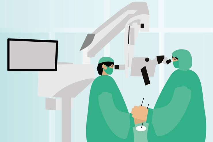
Otolaryngology (ENT) Microscopy Solutions
Otolaryngology (ENT) microsurgery requires top-class imaging quality and visualization. Microscopes for otolaryngology allow specialists to carry out complex and minimally invasive ENT surgical procedures with a high level of precision to enable the best possible clinical outcomes.
Leica Microsystems offers advanced solutions to support ENT specialists in microsurgical treatment. Our innovative microscopy technologies provide enhanced depth of field, precise positioning, and great maneuverability.
ENT surgical microscopes from Leica Microsystems provide a diverse and adaptable solution to ENT specialists for a wide range of otolaryngologic microsurgical procedures. They can be customized and integrated with other devices, including binoculars, objective lenses, handles, stands, and more.
Contact our imaging specialists for expert advice to help you select the right ENT microscope for your requirements and budget.

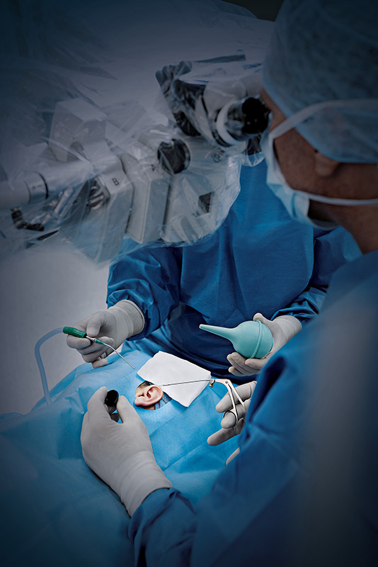
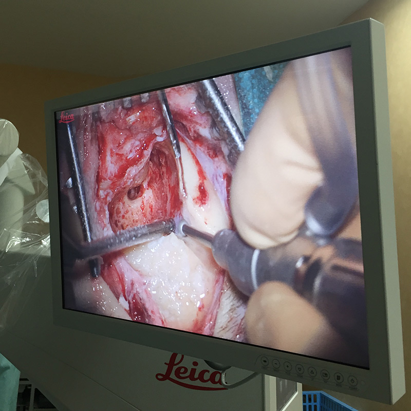
Visualize deep cavities thoroughly also during non-surgical procedures like placement of transtympanic aerator or foreign body removal.

Premium apochromatic optics and tailored illumination provide a crisp, true-color image even at the bottom of the deep, narrow channels typical of ENT surgery.

ENT procedures require different operating positions which is why our microscopes can be maneuvered and positioned with ease, while you retain an upright posture.

Select from an overhead or floor stand according to your OR setup, then customize the optics so you and your assistant can achieve comfortable working positions.
Applications of ENT surgical microscopy
ENT specialists rely on ENT microscopes to visualize the surgical site and enable them to perform a wide range of surgical procedures with a deeper and more detailed view. Some of the most common otolaryngology procedures require the use of an ENT surgical microscope, including:
- Neurinoma surgery
- Otosclerosis surgery
- Schwanoma surgery
- Cholesteatoma surgery
- Cochlear implant surgery
- Stapedectomy
- Tympanoplasty
- Laringoplasty
- Myringoplasty
Challenges of ENT surgical microscopy
One of the major challenges of ENT surgery is maintaining sufficient illumination while navigating complex anatomical structures and deep, narrow cavities. Proper illumination during ENT surgery is crucial for accurate location and access of the surgical site while minimizing iatrogenic injury to nearby structures and tissues.
Leica ENT microscopes provide bright xenon or LED illumination which allows light to penetrate even narrow cavities for a precise visualization. Many also feature BrightCare Plus which automatically adjusts light intensity to protect tissue.
BrightCare Plus – Light Intensity Control
Our innovative BrightCare Plus technology automatically adapts light intensity according to the microscopes’ working distance to prevent damage to sensitive tissues. The BrightCare Plus feature allows maximum illumination at long working distances. As working distance decreases, the light intensity is reduced automatically, diminishing the risk of tissue damage and burns (up to 60% reduction of light intensity). Conversely, the light intensity increases proportionally with working distance.
BrightCare Plus is available in the M530 OHX Microscope and PROvido for ENT surgery.
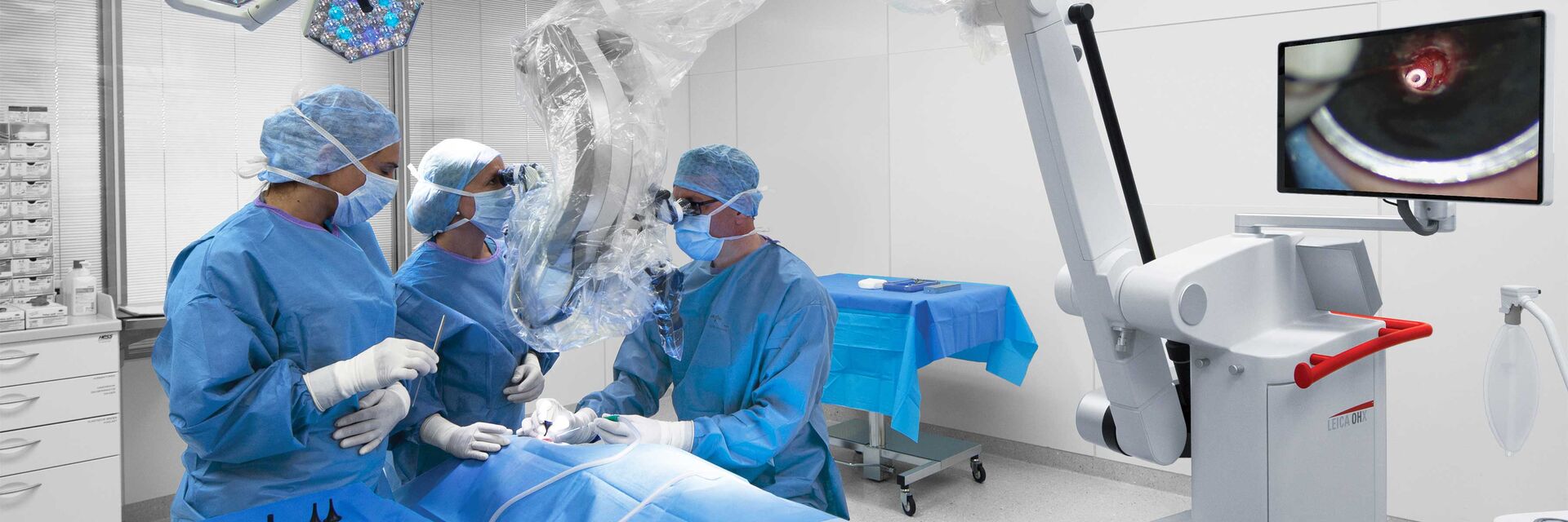
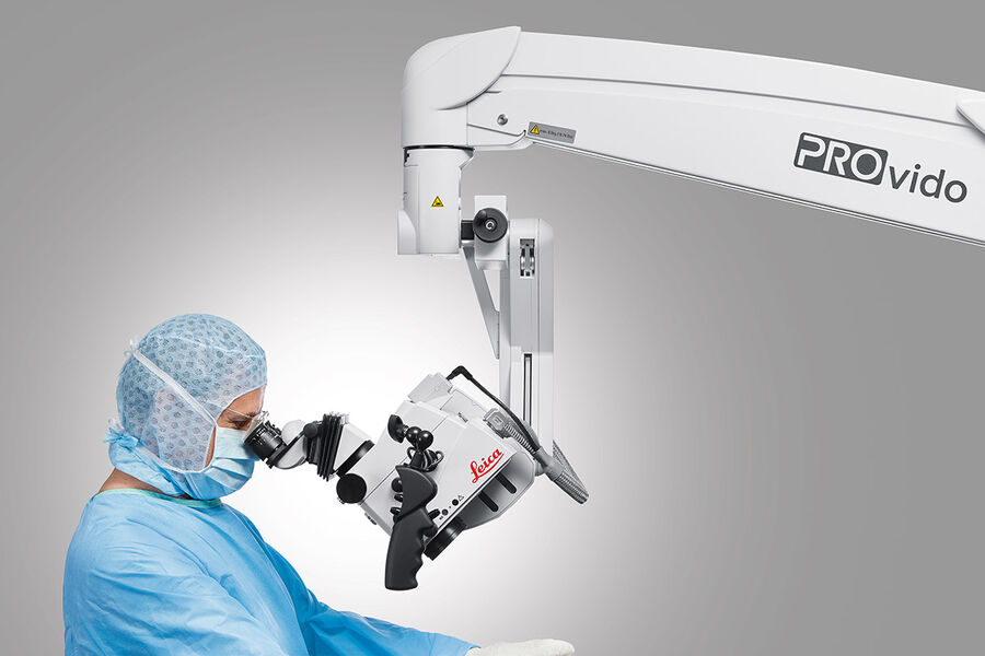
Adaptability of Leica ENT surgical microscopes
During ENT surgical procedures, medical professionals typically work while seated and are closely positioned to the operating field. Alongside optical quality the adaptability of the microscope, as well as intuitive operation are therefore some of the most important features to consider when choosing an ENT microscope.
Using an ENT microscope with a small optics carrier allows the ENT specialist to be in close proximity to the patient, while maintaining maximum hand-eye coordination due to significant gains in working distance. This can contribute to a more ergonomic working position for the ENT specialist, as well as the side assistant. A comfortable working position can contribute finally to the efficiency of procedure.
Adaptable ENT microscopes should offer:
- Positioning freedom & easy maneuverability allowing smooth movement of the microscope with one hand.
- Adaptable binoculars that can be positioned and angled in order to fit specific operating requirements.
- Intuitive and easy-to-reach microscope controls, including programmable footswitch and handles.
- Resistance to vibrations and sturdy design
Integration of ENT microscopes with other devices
Leica ENT surgical microscopes can be integrated with other devices to meet a wide spectrum of requirements in clinical use and teaching. We offer an assortment of product accessories to meet the needs and preferences of ENT specialists for increased efficiency and smooth workflows. Some examples of accessories and devices that can be used to customize our microscopes for otolaryngology include the following:
- Binoculars and objective lenses
- Single or dual handles
- Imaging and recording systems
- Stand-mounted monitors of various sizes
- Side observers
- Micromanipulator for CO2 laser surgery
- Fluorescence modules
Important considerations for selecting an ENT surgical microscope
Some of the key factors that should be considered when selecting an ENT microscope for surgical procedures include the following:
- Illumination to work in narrow and deep cavities
- High magnification that permits the ENT specialist to access and target small, delicate structures with high precision
- Depth of field that provides a clear, three-dimensional view of the surgical field
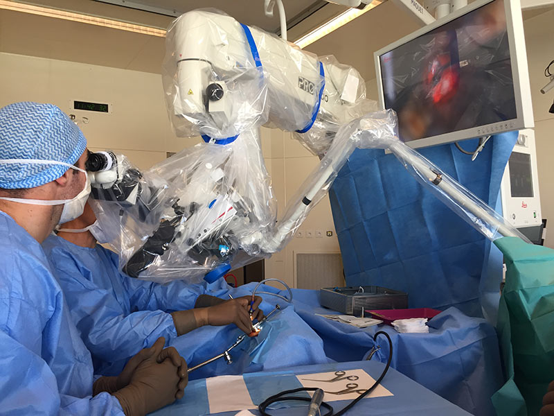
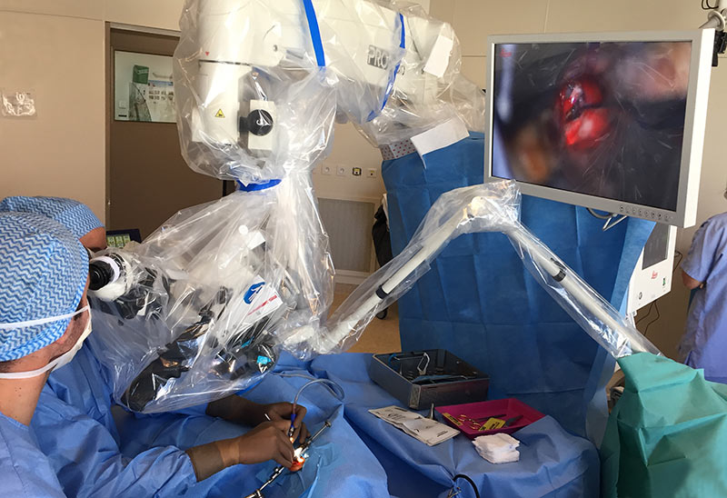
PROvido multidisciplinary surgical microscope
The advantages of using PROvido in ENT surgery include the following:
- Fully focused view into deep and narrow cavities for precise surgical procedures.
- High Maneuverability for smooth microscope movements during surgery, e.g. with two fingers or one hand.
- Adjustable and ergonomic optics for more working comfort
ENT microscope solutions from Leica Microsystems
Leica Microsystems offers a wide range of solutions for ENT specialists to achieve optimal results in both clinical to surgical settings. Our portfolio of products for ENT procedures includes the following features:
- Premium apochromatic optics and brilliant light for high-resolution, true-color images of anatomical structures.
- Maneuverability and mobility allowing smooth movement and easy positioning of the ENT microscope for different operating positions.
- Overhead or floor stands and customized optics for ergonomic and comfortable working positions.
Other useful accessories for ENT surgical microscopes include:
- HDC 100 Integrated Camera to capture high-definition video or still images of surgical procedures for image sharing by teams of specialists and for teaching purposes.
- Evolution4K Recorder from MedXchange to capture 4K UHD video and 4K still images and record to external hard drives or flash drives.
- Integrated Screens of different sizes for optimal visualization during ENT procedures.


 or Minor’s syndrome.")