Neurovascular Surgery Solutions
For precise, real-time insights into cerebral anatomy and blood flow during neurovascular procedures, Leica surgical microscopes with fluorescence imaging and 3D augmented reality provide full depth perception to neurosurgeons treating aneurysms and arteriovenous malformations (AVMs) and performing bypass surgeries.
Contact a local imaging specialist for expert advice on the right Neurovascular Surgery Solutions microscope for your needs and budget.


What methods ensure precise surgical visualization?
Neurosurgeons can clearly distinguish between critical vessels and surrounding areas, thanks to the crisp images and natural colors provided by Leica surgical microscopes. FusionOptics technology delivers high-resolution and high depth of field in one image, allowing surgeons to see an optimal spatial image and a significantly expanded area in full focus.

How do neurosurgeons ensure vessel patency intraoperatively?
To prevent complications during neurovascular surgery such as inadequate blood flow or vessel occlusion, visualization the blood flow is essential. For surgeons to determine vessel patency, the FL800 from Leica Microsystems provides intra-operative fluorescence-aided video angiography for real-time visualization of blood flow.

How to visualize blood flow and anatomy simultaneously?
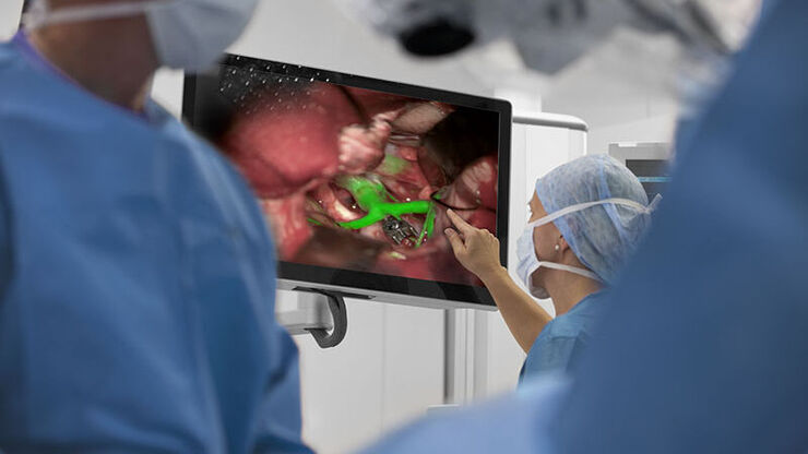
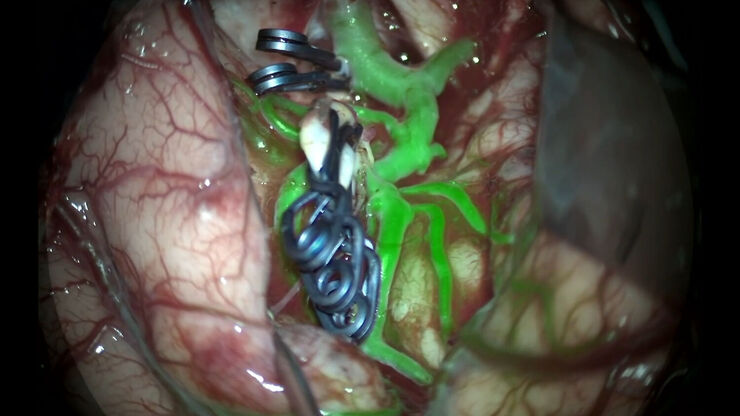
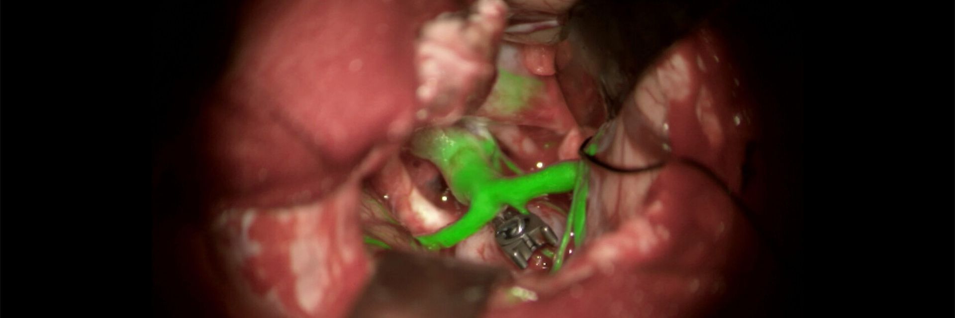
Simultaneous visualization of anatomical and vascular structures helps surgeons make accurate assessments during neurovascular surgery. With the GLOW800 AR fluorescence application and ICG dye, surgeons can observe cerebral anatomy in natural color, augmented by real-time vascular flow in 3D resolution.

Is it possible to minimize surgical interruptions?
Integrating natural white-light microscope images with near-infrared images in one augmented real-time view allows surgeons to see all necessary details. With Leica surgical microscopes, surgeons no longer need to switch between the natural microscope image and a flat black-and-white near-infrared (NIR) video, maintaining a smooth workflow and reducing strain.
Why should you choose a Leica surgical microscope for your neurovascular surgeries?
Cleverly Enhanced Optical Visualization
To perform neurovascular procedures like aneurysm clippings, surgeons using Leica surgical microscopes have a clearer view of the surgical field. FusionOptics technology brings together an enhanced depth of field with high resolution. Plus, Small Angle Illumination (SAI) ensures a shadow-free view into deep cavities.
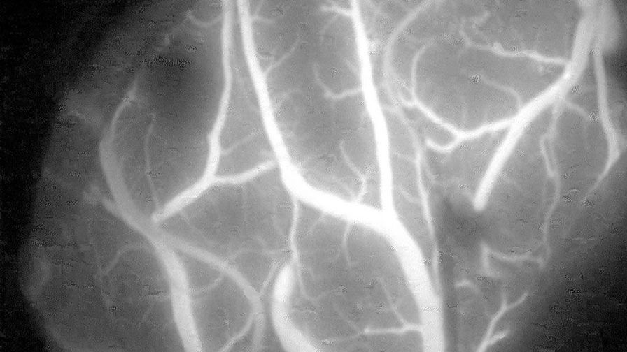
Intra-operative Fluorescence Imaging
The FL800 provides, intra-operative fluorescence-aided video angiography, allowing surgeons to determine the patency of vessels. This innovative neurovascular imaging technology uses Indocyanine green (ICG) fluorescence dye and near infrared cameras to display blood flow directly through the neurosurgical microscope.

Anatomy and Blood Flow in One Augmented Digital View
Surgeons are provided with simultaneous white-light and real-time fluorescent views of the blood flow with the GLOW800 AR fluorescence and the ICG fluorescence dye. This Leica innovation offers surgeons a single augmented image and enhanced precision, eliminating the need to switch between views.
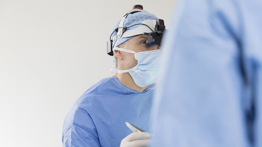
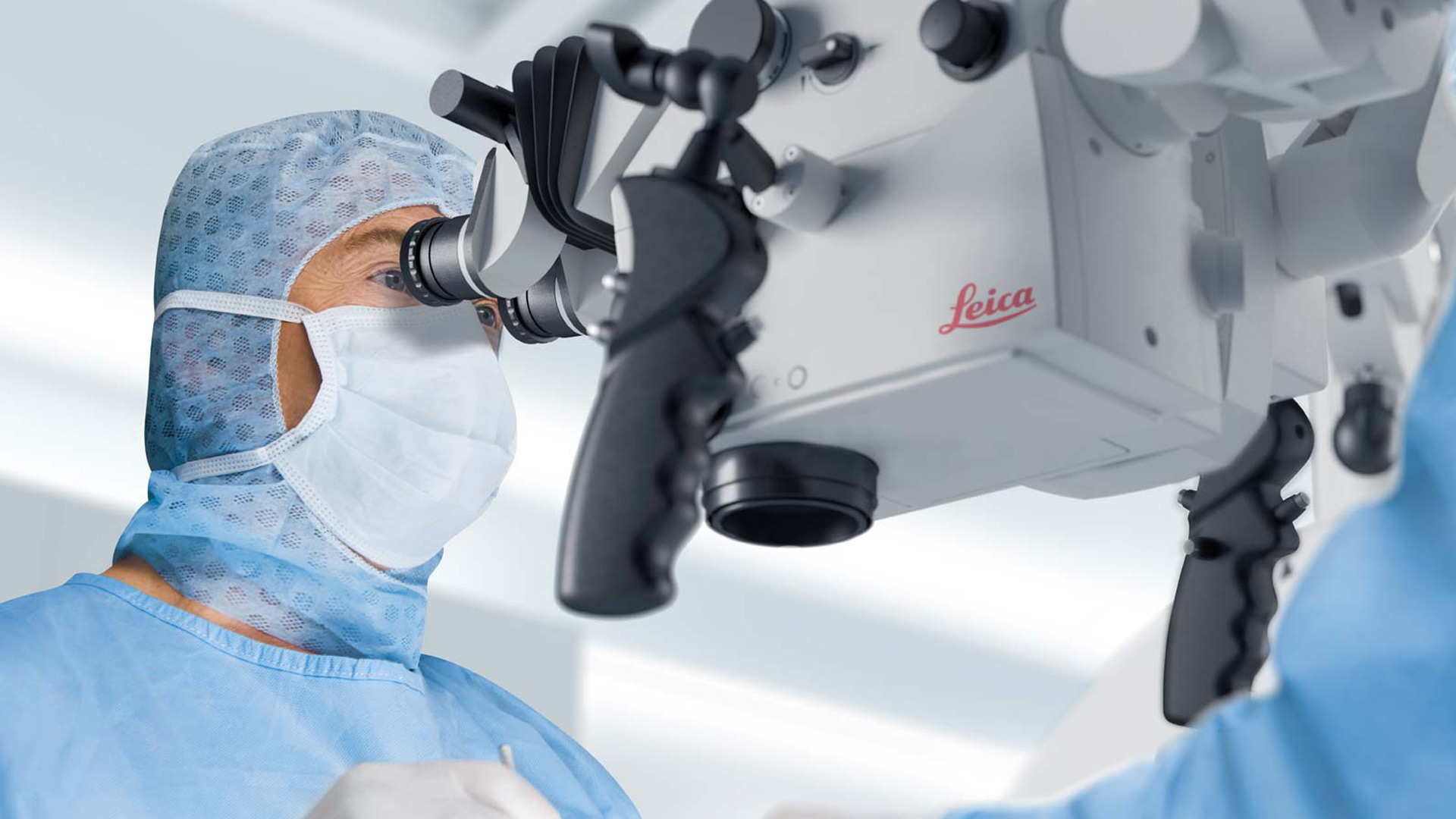
Visualization Freedom
Leica provides a range of visualization options to ensure that surgeons have freedom of movement and can collaborate effectively with their OR colleagues. The Evolved ARveo 8 microscope offers three interchangeable viewing options: traditional oculars, heads-up 2D/3D monitors, or the MyVeo surgical visualization headset. MyVeo frees surgeons from the microscope by unifying essential clinical data right in front of their eyes.
Now we actually can stay oriented in our 3D environment while seeing the blood vessels glow up. So, this was, to me at least as a neurovascular surgeon, a big advantage and a big achievement!
With GLOW800 augmented reality fluorescence, it is possible to see the angiographic picture in the microscope directly during neurovascular surgery, with an optimal depth perception. There is no need to recall and try to reconcile the black and white blood flow video with the natural anatomical view.
A unified view of the cerebrovascular region
The FL800 Intra-Operative Fluorescence system enables surgeons to visualize blood flow using near-infrared (NIR) imaging and indocyanine green (ICG) dye to determine the patency of the vessels. The GLOW800 Augmented Reality Fluorescence Application enhances this by integrating real-time vascular flow with natural color views of cerebral anatomy. This provides surgeons with a 3D augmented reality experience in real-time, eliminating the need to reconcile black-and-white blood flow video with the natural anatomical view.


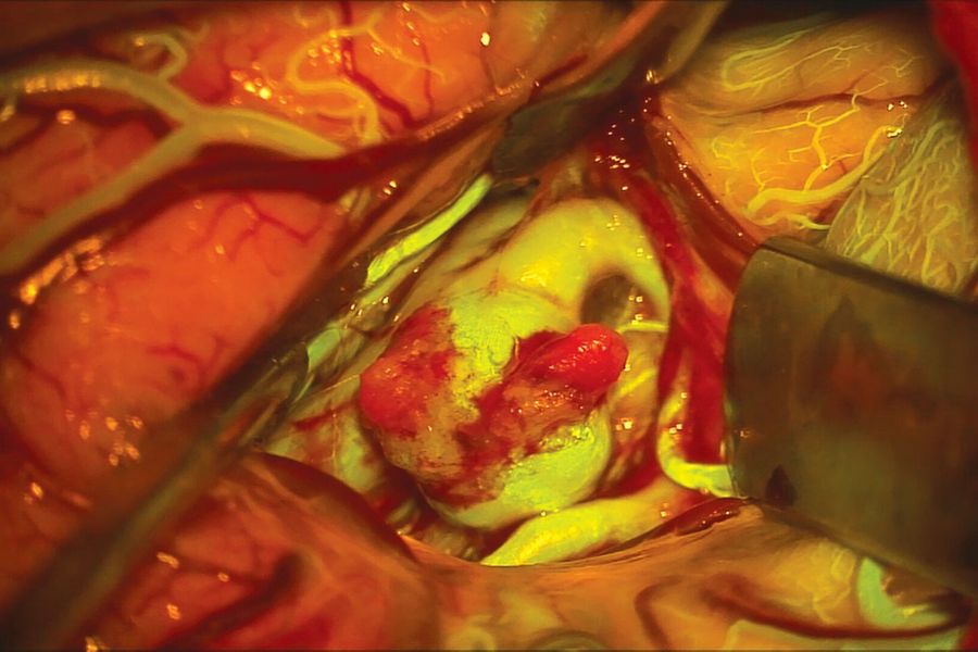
Images courtesy of Cleopatra Charalampaki, MD, PhD, Professor of Neurosurgery, Department of Neurosurgery, Cologne Medical Center, Germany.
Neurovascular fluorescence
The FL560 fluorescence module, used with fluorescein, allows surgeons to simultaneously view anatomical structures and cerebrovascular blood flow, clearly differentiated and with high contrast. It enables simultaneous, real-time observation of both non-fluorescent tissue and fluorescent areas. The FL560 fluorescence module provides one complete, high-resolution, high-contrast image for a crisp anatomical view and clearly delineated vascular flow, even in tiny vessels.
Frequently asked questions Neurovascular surgery solutions
Neurovascular surgery microscopes are specialized medical devices designed to provide precise visualization and imaging during surgeries like aneurysm clipping, arteriovenous malformations (AVMs), and bypass procedures.
Surgical microscopes and fluorescence imaging can be utilized in aneurysm procedures involving the Anterior Cerebral Artery and Anterior Communicating Artery, bypass procedures for conditions such as Moyamoya disease, and treatments for arteriovenous malformations (AVMs). Additionally, these technologies are applicable to all anastomosis surgeries, including those in plastic and reconstructive surgery.
Fluorescence angiography uses ICG dye and specialized fluorescence neurosurgery systems to visualize blood flow in real-time. Leica Microsystems combines this with GLOW800 AR fluorescence for simultaneous anatomical and vascular views.
AR fluorescence in neurosurgery integrates real-time anatomical and vascular imaging into a single augmented view, enabling surgeons to make accurate intraoperative decisions without switching imaging modes.
The choice depends on the surgeon's needs and habits. Options include the FL800 in black-and-white near-infrared (NIR) or the GLOW800 AR, which offers both black-and-white and pseudocolor imaging, combining vascular flow and anatomical visualization. Carefully consider your surgical requirements, including the type of fluorescence, imaging integration, and ergonomic adaptability.
Depending on your preference for standard fluorescence (FL800) or advanced visualization (GLOW800 AR), as well as the configuration and customization, the price of a neurovascular surgery microscope can vary. Additionally, the use of fluorescein with the FL560 fluorescence module is a low-cost alternative for neurovascular imaging.
Leica Microsystems offers on-site demonstrations, and clinical applications support to help surgeons and OR teams maximize the use of their neurovascular surgery microscopes and fluorescence applications.
Yes, while primarily designed for neurovascular surgery/neurosurgery, these microscopes can be adapted for other surgical disciplines requiring precise visualization, such as ENT and spinal surgeries.