
Clinical Laboratories
Clinical Laboratories
What are you interested in? Show subnavigation
Clinical Laboratories

Products Show subnavigation
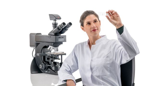
Visoria B
Experience enhanced efficiency and comfort in your daily microscopy routine. The Visoria B laboratory microscope is for applications performed in life science and clinical laboratories.
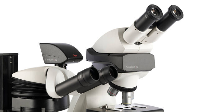
Flexacam c5 & i5
With Flexacam microscope cameras, you can see the fine sample details you need to see with precise colors and high resolution. Viewing, documenting, and sharing microscope images of your samples the...
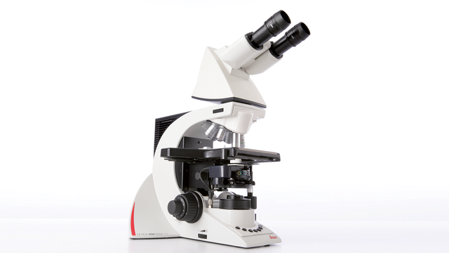
Leica DM3000 & DM3000 LED
Uniquely Ergonomic System Microscopes With Intelligent Automation
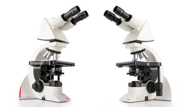
DM1000
Uniquely Ergonomic System Microscope
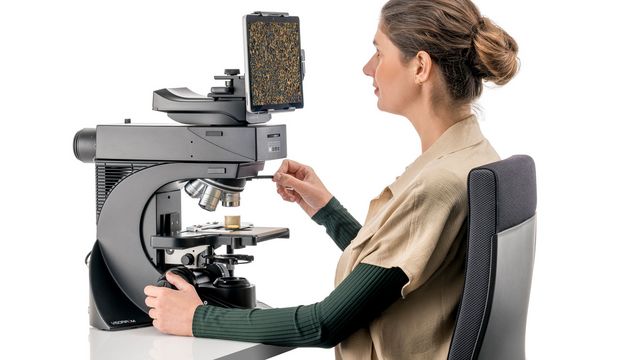
Enersight
What if you could inspect, compare, measure and share with one intuitive interface? Introducing Enersight, a powerful software platform for microscopy.
Leica Science Lab Show subnavigation
Read our latest articles about Forensic Science and Imaging Equipment
The knowledge portal of Leica Microsystems offers scientific research and teaching material on the subjects of microscopy. The content is designed to support beginners, experienced practitioners and scientists alike in their everyday work and experiments.
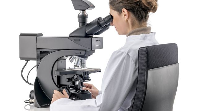
Factors to Consider when Selecting Clinical Microscopes
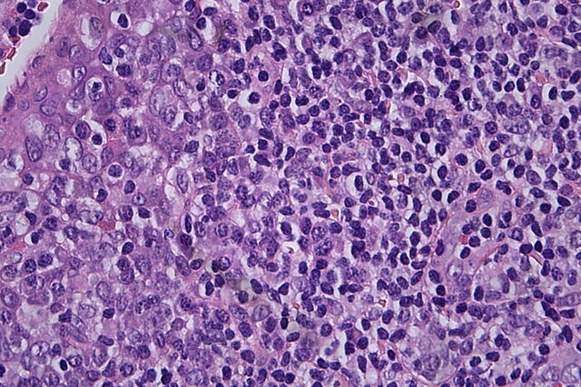
Clinical Microscopy: Considerations on Camera Selection
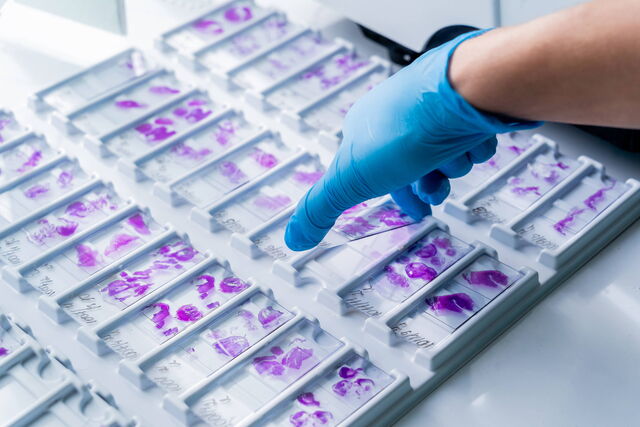
H&E Staining in Microscopy

The Time to Diagnosis is Crucial in Clinical Pathology
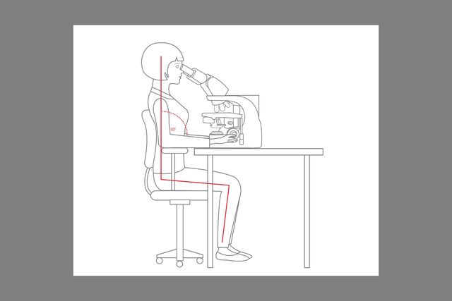
Perform Microscopy Analysis for Pathology Ergonomically and Efficiently
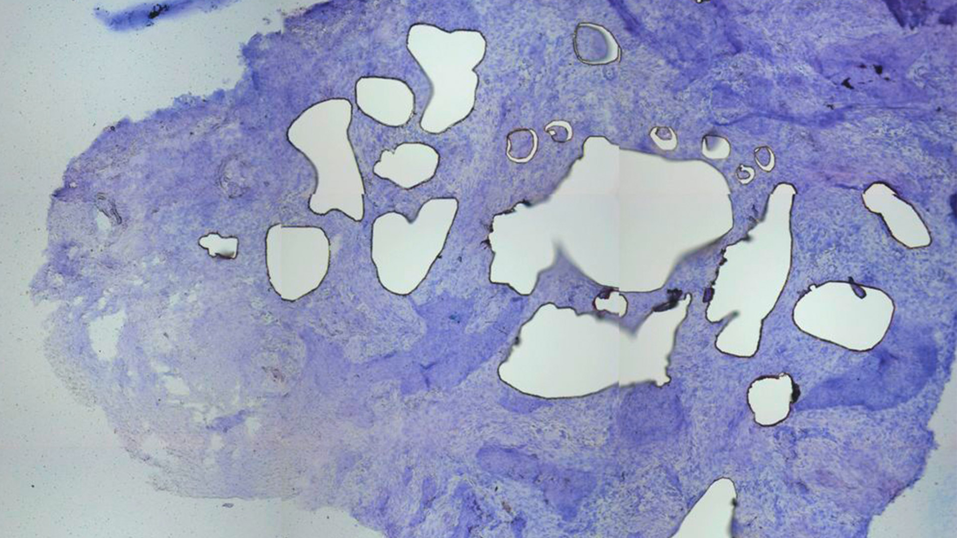

What are challenges concerning microscopy for pathology?
Pathologists need to prepare specimen slides for microscopic examination and ensure the accuracy and reliability of diagnoses. This work is often performed under strict time constraints, as results are urgently needed to support clinical decision making in hospitals and laboratories. Leica microscopes help pathologists achieve efficient and reliable specimen processing.

How is microscopy used for pathology?
Microscopy refers to any type of examination in the pathology lab workflow that is performed with a microscope. Examples are checking the quality of specimen staining as well as the examination and documentation of specimens. Leica microscopes help users reach pathological diagnoses efficiently and comfortably.

Why do pathologists use microscopes?
Microscopes enable pathologists to see abnormalities that they would not be able to see with just their eyes, whether they are fine structures, subtle differences in color, or the number of specific cells in a specimen. Leica microscopes offer various contrast methods that can make even more details visible.

What is the difference between clinical and anatomic pathology?
Clinical and anatomic pathology concern different specimens. Clinical includes chemical, molecular, immune-, and hematopathology, as well as medical microbiology. Anatomic focuses on histopathological examinations of tissues or cells used in surgical, cyto-, forensic, and dermatopathology. Pathologists benefit from a Leica microscope that allows them to achieve efficient diagnoses.
Why use Leica microscopes for clinical laboratory and pathology work?
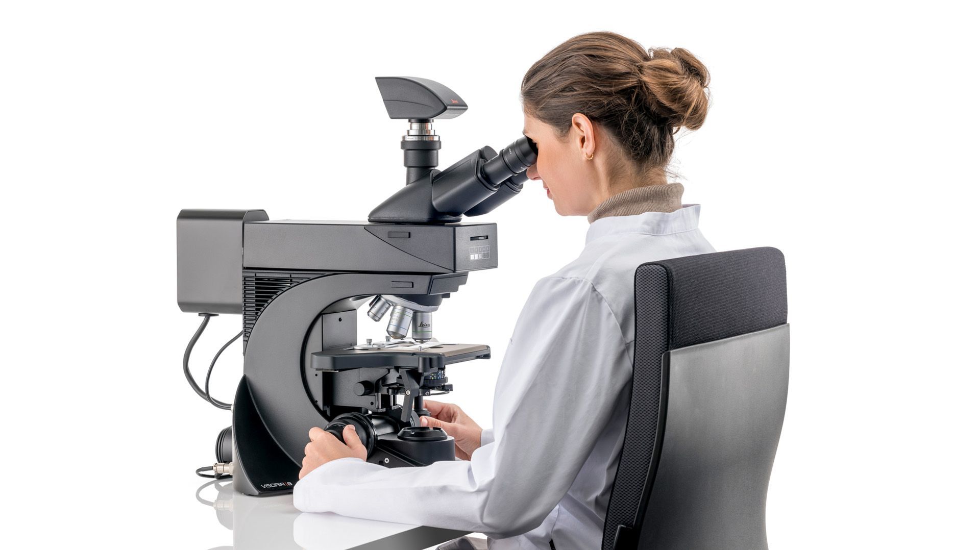
Work in comfort
With Leica microscopes, you maintain a proper posture, reducing the risk of neck and back strain during long hours at the microscope. Work comfortably with aligned shoulders and spine. Attain ergonomic hand and arm positioning due to the symmetrical layout of the focus stage control knobs and ergonomic accessories.
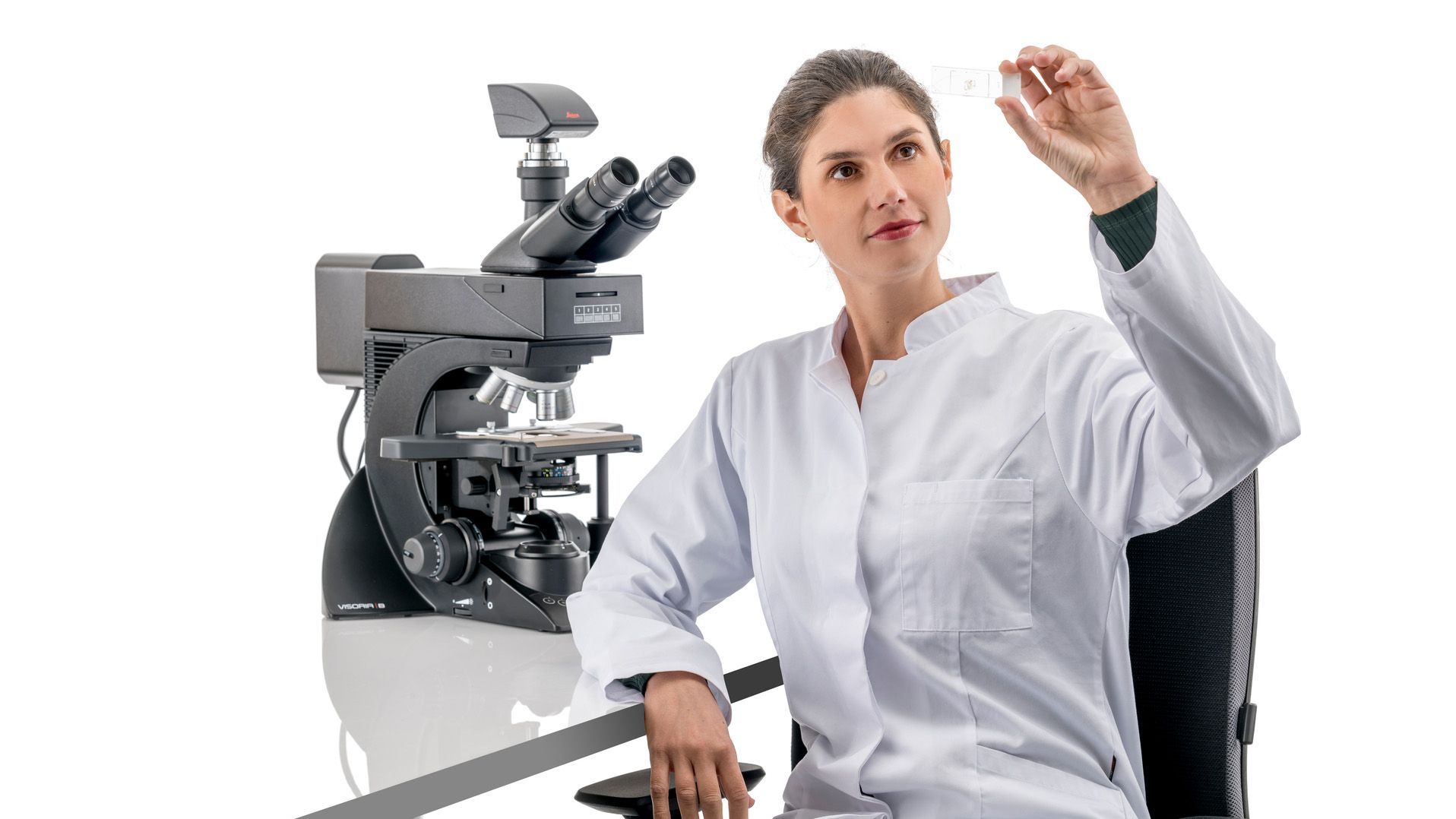
Made for clinical laboratories
Leica microscopes reveal sub-cellular structures in tissues and specimens with various contrast methods, such as brightfield, darkfield, phase contrast, DIC, and fluorescence. For special diagnostic requirements, Leica microscopes are certified for in-vitro diagnostics ( IVD ).
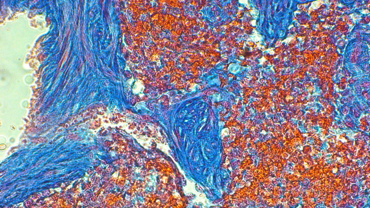
High-quality images
Leica optics deliver images with high contrast and resolution and clear fluorescence signals. These high-quality images enable you to confidently visualize fine specimen details even in low light. There is a broad portfolio of Leica objectives which supports reliable and accurate microscopy for clinical laboratories.
Customer experiences with a DM3000 microscope for clinical applications
Trudi de Jong and Marianne Noordanus, both from Rotterdam, describe how a DM3000 microscope helps them to perform their clinical microscope work more comfortably and efficiently.
Courtesy of:
Trudi de Jong, Erasmus MC academic hospital Rotterdam, the Netherlands, Hematology
Marianne Noordanus, Star-MDC, Medisch Diagnostisch Centrum, Rotterdam (The Netherlands) Microbiology
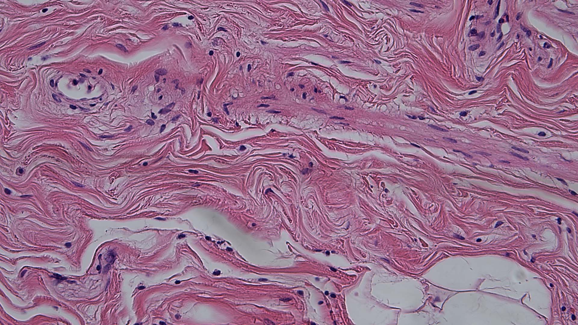

Frequently Asked Questions Pathology
Cameras are used for documentation, the displaying of images on a larger screen, and showing live images for discussion among colleagues on tumor boards. They also help pathologists with reporting results, especially if the camera comes with software that allows annotating and archiving images in the laboratory or hospital information system.
If pathologists need to see structures and shades of color in stained specimens, they will look at specimens in brightfield. If they need to identify the structures of cells and tissues in unstained specimens, they will use phase contrast.
Pathologists can work with color or monochrome cameras. Color cameras are ideally suited for pathology applications as they can visualize minute differences in staining and give pathologists a wealth of information about the specimen. Monochrome cameras are ideally suited for fluorescence applications, such as FISH (fluorescence in situ hybridization).


